 Deca Head Microscope
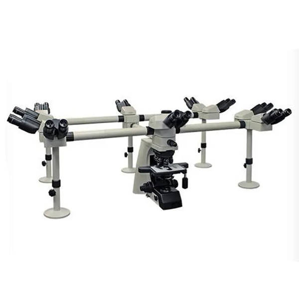
A Deca Head Microscope supports group learning and shared analysis at one workstation. This system features ten viewing heads arranged around a single optical body. You and your team view the same specimen simultaneously. This setup suits medical colleges, dental colleges, training centers, and research labs.
The deca-head microscope design focuses on clarity, comfort, and coordination. Each user receives a direct view through a personal eyepiece. No screen delay occurs. No interpretation gap appears between users. Faculty guide students in real time. Students follow every step without shifting seats.
What a Deca Head Microscope Means for Your Lab
You train groups daily. You review slides with interns. You explain tissue structure to a full batch. This microscope solves space and time limits. Ten people work together on one sample. Discussion stays focused. Learning speed improves.
Common Use Environments
Medical colleges
Dental colleges
Pathology labs
Histology training centers
Research institutions
Teaching hospitals
Core Optical Structure
A Deca Head Microscope uses an infinity corrected optical system. Parallel light travels through the optical path. Each head receives the same image quality. Image scale stays consistent across all viewing stations.
Main Optical Elements
Infinity corrected objectives
Wide field eyepieces
Multi port head assembly
Central optical body
This structure reduces image variation between users. Faculty see the same details as students. Demonstration remains clear.
Viewing Heads and Ergonomics
Ten heads surround the central body. One head supports the instructor. Nine heads support learners. Each head includes a binocular tube. Head angles support long sessions.
Typical Head Features
Binocular viewing
Inclined viewing angle around 30 degrees
Individual diopter adjustment
Comfortable eye spacing
This arrangement reduces neck strain. Users maintain posture during long classes.
Eyepieces and Field of View
Wide field eyepieces support group teaching. A common specification uses EWF 10x eyepieces with a 22 mm field. This wide field shows more sample area. Users grasp context faster.
Eyepiece Benefits
Large viewing circle
Uniform brightness across field
Comfort for extended viewing
Faculty point out structures without repeated repositioning.
Objective System
Objective lenses define magnification and resolution. A Deca Head Microscope uses an infinity plan objective set. Common magnifications include 4x, 10x, 40x, and 100x oil.
Objective Advantages
Flat field across view
Consistent scale for all heads
Smooth transition between magnifications
Students learn tissue comparison by switching powers during the same session.
Nosepiece Design
A quadruple nosepiece holds four objectives. Rotation remains smooth. Click stops align lenses correctly. Instructors change magnification during explanation.
Nosepiece Traits
Metal construction
Positive click stops
Easy access for cleaning
This part supports daily classroom use.
Stage and Specimen Handling
The mechanical stage supports precise slide movement. A double layer stage adds stability. Slide controls move smoothly along X and Y axes.
Stage Specifications
Stage size around 180 mm by 160 mm
Slide holder with firm grip
Smooth coaxial movement
You position a structure once. All users see the same area.
Focusing Mechanism
Coaxial coarse and fine focus knobs support clear image control. Fine focus allows minute adjustment. Coarse focus brings the image into view quickly.
Focus System Benefits
Balanced tension
Smooth rotation
Stable focus under group use
Instructors focus once. Students follow without refocusing.
Condenser System
An Abbe condenser with NA 1.25 supports contrast control. Height adjustment aligns light path. Iris diaphragm manages contrast and depth.
Condenser Features
Centering adjustment
Smooth height control
High numerical aperture
This system supports stained and unstained samples.
Light Source System
This microscope uses a built in light source. Options include halogen or LED systems. Brightness control supports specimen type and stain density.
Light System Traits
Stable output
Low heat near stage
Long operating life
Users adjust brightness using a dedicated control.
Power Requirements
Standard power input uses 230 V AC. Voltage compatibility suits lab infrastructure in India and similar regions. Internal protection supports stable operation.
Installation Requirements
Standard power outlet
Stable table
Clean environment
Setup time stays short.
Teaching Workflow with a Deca Head Microscope
A typical teaching session follows a simple pattern.
The instructor prepares a slide.
The slide moves onto the stage.
Focus occurs once.
All learners view together.
Discussion flows naturally. The instructor points through verbal guidance. Students ask questions while viewing the same structure.
This process saves time. No queue forms. No repeated alignment occurs.
Applications in Medical Education
Medical students study histology and pathology daily. Group viewing improves understanding of tissue patterns. Faculty explain cell layers and structures live.
Common Subjects
Histology
Pathology
Microbiology
Hematology
Students link theory with visuals during the session.
Applications in Dental Education
Dental students analyze oral tissues and tooth sections. Shared viewing helps faculty explain enamel, dentin, pulp, and periodontal structures.
Typical Use Cases
Oral pathology
Dental histology
Prosthodontic material study
Group learning reduces equipment duplication.
Applications in Research Labs
Research teams review samples together. Principal investigators guide junior researchers. Consensus forms faster.
Research Benefits
Shared interpretation
Faster decision making
Reduced sample handling
This system supports collaboration.
Build and Stability
A Deca Head Microscope requires a strong base. The stand supports the weight of ten heads. Anti vibration feet improve stability.
Structural Traits
Metal stand
Wide footprint
Balanced arm design
This structure supports long term institutional use.
Maintenance Practices
Daily care keeps the microscope functional.
Clean eyepieces after sessions.
Cover the microscope when idle.
Switch off power after use.
Clean objectives using proper lens paper.
Annual servicing supports consistent operation.
Space Planning for Your Lab
This microscope needs a large table. Each user requires seating space. Plan circular seating.
Recommended Setup
Central table
Ten stools
Clear walkways
Controlled lighting
Proper planning improves session flow.
Selection Checklist for Buyers
Before purchase, review these points.
Number of heads required
Objective magnifications included
Light source type
Service availability
Warranty terms
Match features with teaching load.
Advantages for Institutions
Institutions reduce equipment count. One microscope replaces multiple units. Faculty manage large groups without rotation.
Operational Benefits
Lower setup time
Unified teaching
Better engagement
Administrative planning improves.
Training Outcomes
Students learn faster through shared visuals. Questions arise at the right moment. Faculty address doubts immediately.
Observed Outcomes
Higher attention
Clear concept building
Reduced repetition
Teaching quality improves.
Safety and Handling
Follow basic safety steps.
Avoid touching lenses with fingers.
Keep liquids away from power parts.
Handle slides carefully.
These steps protect users and equipment.
Why a Deca Head Microscope Fits Academic Growth
As batch size grows, teaching tools must scale. This microscope supports expansion without adding multiple units. Faculty maintain teaching style while reaching more learners.
This system suits institutions aiming for efficient lab teaching.
Deca Head Microscope Summary
A Deca Head Microscope supports ten users viewing one specimen simultaneously. This design suits education and collaborative research. Optical consistency across heads ensures shared understanding. Mechanical stability supports daily use. Institutions benefit from time savings and improved group instruction.
You gain a structured teaching workflow. Students gain clarity during lessons. Faculty guide without interruption. This microscope stands as a practical solution for group microscopy training.
Independent SEO consultant specializing in laboratory and medical equipment brands, focused on ranking high-intent keywords, strengthening authority, and driving sustainable organic growth through ethical, data-driven SEO.