A microscope is a tool used to see objects too small for naked eyes. Human eyes fail to see cells, bacteria, fibers, or fine surface details. A microscope solves this problem. Lenses inside the microscope enlarge small objects by bending light. The enlarged image reaches your eyes with more detail. Doctors use a microscope to study blood samples and tissues. Scientists use a microscope to study cells and microorganisms. Students use a microscope in schools and colleges to learn biology and chemistry. Factories use a microscope to inspect tiny defects in materials. A microscope exists to show hidden structures clearly and simply.
Microscopes have several types. The main types are:
- Compound Microscope
- Binocular microscope
- Digital Microscope
- Stereo Microscope
- Fluorescence Microscope
1. Compound Microscope
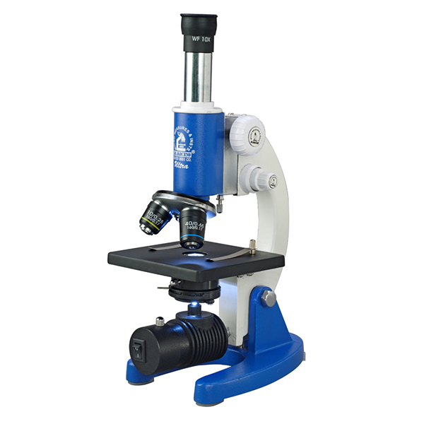
A compound microscope lets you see tiny objects on a glass slide. It has two lenses. The objective lens sits near the object and forms a small image. The eyepiece lens enlarges this image for your eyes. It gives higher magnification than simple microscopes and reduces color errors. Doctors, students, and scientists use it to study cells, bacteria, and tissues
Diagram:

2. Binocular microscope

A binocular microscope is a type of microscope with two eyepieces. It allows you to look with both eyes at the same time. This makes viewing more comfortable and reduces eye strain. The lenses work like a compound microscope, with an objective lens near the specimen and an eyepiece lens for magnification. It is used to study cells, tissues, and small objects in labs.
Diagram:

3. Digital Microscope
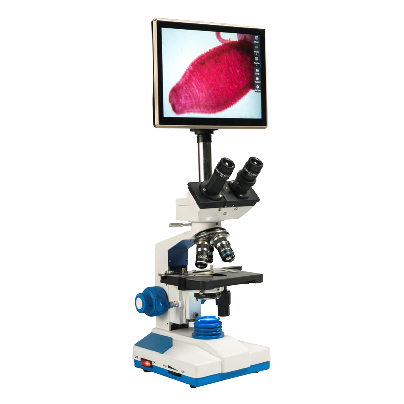
A digital microscope is a microscope that shows the magnified image on a computer or screen. It uses a camera instead of traditional eyepieces. You can capture photos and videos of the specimen. It allows multiple people to view the object at the same time. It is used in schools, labs, and industries to study cells, insects, materials, and small defects in objects.
Diagram:
4. Stereo Microscope
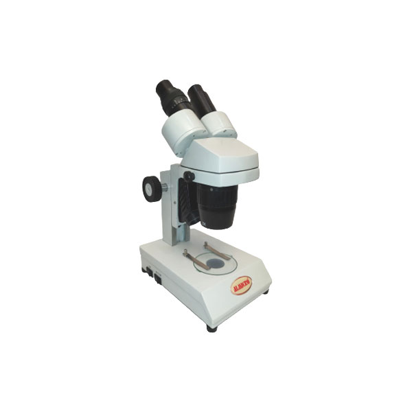
A stereo microscope is a low-power microscope that shows objects in 3D. It has two eyepieces and two separate optical paths. This creates a three-dimensional view of the specimen. It is used to study insects, plants, circuit boards, and small mechanical parts. It does not give very high magnification but helps see shapes and surfaces clearly. It is useful for detailed work and inspection.
Diagram:

5. Fluorescence Microscope
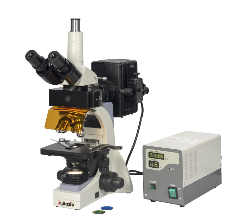
A fluorescence microscope is a microscope that uses special light to make certain parts of a specimen glow. It highlights specific structures like cells, proteins, or tissues. Fluorescent dyes or markers are used to show these parts clearly. Scientists use it to study cells, bacteria, and biological processes. It helps see details that are invisible under normal light.
Diagram:
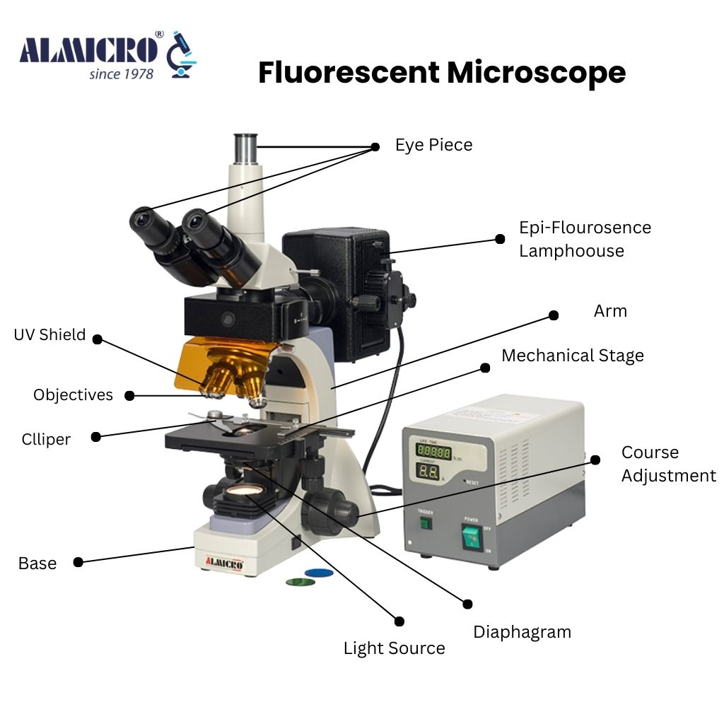
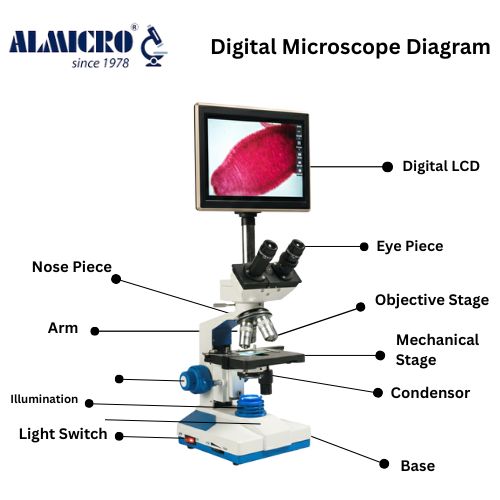
List the main components one by one in Every Microscope. For example:
- Eyepiece – Lens you look through, magnifies the image.
- Objective lenses – Close to specimen, create first magnified image.
- Nosepiece – Holds objective lenses, rotates to change magnification.
- Stage – Flat platform to place the slide, holds specimen.
- Stage clips – Keep the slide in place.
- Light source – Shines light through the specimen to see details.
- Diaphragm – Controls the amount of light passing through the specimen.
- Coarse focus knob – Moves stage or lenses quickly for general focus.
- Fine focus knob – Adjusts focus slowly for clear, sharp image.
- Arm – Connects base to top, used for holding the microscope.
- Base – Supports the microscope and keeps it stable.
- Condenser – Focuses light onto the specimen for better clarity.
History of the Microscope
1. Early Lenses and Magnifying Glasses
People wanted to see tiny objects for centuries. In the 13th to 16th centuries, simple magnifying glasses were used. These lenses made objects look bigger but showed little detail. They were the first step in the development of microscopes.
2. First Compound Microscope by Zacharias Janssen

In the late 1500s, Zacharias Janssen, a Dutch lens maker, created the first compound microscope. It had two lenses and gave higher magnification than a single lens. Early compound microscopes were not very clear but allowed people to see smaller objects better than before.
3. Improvements by Galileo Galilei
In the early 1600s, Galileo improved the microscope. He made better lenses and showed how multiple lenses could magnify objects more clearly. His work helped scientists understand how to see tiny details.
4. Robert Hooke and the Discovery of Cells
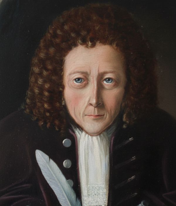
In 1665, Robert Hooke used a compound microscope to study thin slices of cork. He noticed small box-like structures and called them “cells.” This was the first time the word “cell” was used in biology.
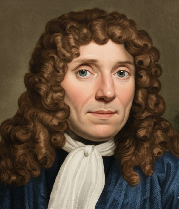
5. Anton van Leeuwenhoek and Microorganisms
In the late 1600s, Anton van Leeuwenhoek built simple microscopes with a single high-quality lens. He looked at water, blood, and tiny organisms. Leeuwenhoek was the first to see bacteria, protozoa, and other microscopic life, opening a new world in biology.
6. 18th and 19th Century Improvements
During the 18th and 19th centuries, achromatic lenses were developed to reduce color distortion. Compound microscopes became more common in laboratories and schools. Stages, focus knobs, and light sources were added to make microscopes easier to use.
7. Modern Microscopes: Electron and Digital
In the 20th century, electron microscopes were developed. Transmission Electron Microscopes (TEM) show internal structures of cells. Scanning Electron Microscopes (SEM) show surface details. Digital microscopes use cameras and screens to display magnified images. Multiple people can view the specimen at the same time.
8. Uses of Microscopes Today
Microscopes are used in many fields. Doctors study blood, tissues, and bacteria. Scientists study cells, microorganisms, and materials. Students use microscopes in schools for biology and chemistry. Industries use them to check small defects in machines, electronics, and products.
The history of the microscope shows the journey from simple lenses to modern digital and electron microscopes. Each improvement helped humans see smaller details, study biology, and explore a world invisible to the naked eye. Microscopes continue to evolve and play a key role in science, medicine, and education.
Applications in Daily Life
Microscopes are used in medicine to study blood, tissues, and bacteria. Scientists study cells, microorganisms, and materials. Students use them in schools for biology and chemistry lessons. Industries use microscopes to inspect tiny defects in machines, electronics, and products. Forensic labs use microscopes to examine evidence in criminal investigations.
Fun Facts and Milestones
- Anton van Leeuwenhoek called bacteria “animalcules.”
- Robert Hooke named cells after monastery rooms.
- Early microscopes could only magnify 20–30 times.
Milestones Timeline
- 13th century – Magnifying glasses appear
- 1590 – Janssen invents first compound microscope
- 1665 – Hooke observes cells
- 1674 – Leeuwenhoek sees bacteria
- 20th century – Electron microscopes
10. Comparison with Modern Technology
17th-century microscopes were tiny and simple. Today’s electron microscopes reveal details at the atomic level. Digital microscopes allow real-time viewing on screens for multiple users.
11. Images and Diagrams Suggestions
- Old vs modern microscope
- Hooke’s cell sketch
- Leeuwenhoek’s simple microscope
12. Quotes
“The microscope is a window to a world invisible to our eyes.”
Hooke and Leeuwenhoek’s own words describe the excitement of seeing tiny life for the first time.
The history of the microscope shows a journey from simple lenses to advanced electron and digital microscopes. Each improvement helped humans see smaller details, explore cells and microorganisms, and improve science and medicine. Microscopes continue to evolve, revealing a world invisible to the naked eye and expanding human knowledge in countless ways.
Fun Facts About Microscopes
- Anton van Leeuwenhoek called bacteria “animalcules” in his letters.
- Robert Hooke named cells after the rooms in a monastery.
- Early microscopes could only magnify objects 20–30 times.
- Some of Leeuwenhoek’s microscopes were less than 3 cm long.
- The word “microscope” comes from Greek words meaning “small” and “to look.”
- Modern electron microscopes can magnify objects over 10 million times.
- Scientists use microscopes to study art, like analyzing pigments in old paintings.
- Microscopes are used in forensic labs to detect tiny evidence in crimes.
- Without microscopes, bacteria, viruses, and tiny cells would remain invisible to humans.
Frequently Asked Questions
A microscope is a scientific instrument used to see and study objects that are too small to be seen with the naked eye. It works by using lenses or other technologies to magnify tiny objects, allowing us to observe details such as cells, bacteria, fibers, and fine surface structures clearly.
A microscope is needed because the human eye cannot see very small objects like cells, bacteria, and fine surface details. It helps scientists and doctors study things that are invisible to the naked eye.
The first compound microscope was invented by Zacharias Janssen in the late 16th century. His invention used two lenses to achieve greater magnification.
Most microscopes include an eyepiece, objective lenses, stage, light source, focus knobs, arm, base, and condenser. These parts work together to produce a clear magnified image.
Robert Hooke discovered and named “cells” after observing cork under a microscope, marking an important milestone in the history of biology.
Anton van Leeuwenhoek was the first to observe bacteria and microorganisms, revealing a hidden world of microscopic life and advancing biological science.
Modern microscopes offer much higher magnification, better clarity, digital displays, and even atomic-level imaging, unlike early microscopes which were simple and low-powered.
Microscopes are used in medicine, education, research, industry, forensics, and material testing to examine tiny details and solve real-world problems.