A microscope is a precision optical or electronic instrument designed to produce magnified images of objects, structures, or organisms that are too small to be observed clearly by the unaided human eye. The term derives from the Ancient Greek words mikrós (small) and skopeîn (to look or examine). Since their invention in the late sixteenth century, microscopes have fundamentally transformed human understanding of biology, medicine, materials science, and numerous other scientific disciplines.
Microscopes operate on a range of physical principles — from the refraction of visible light in classical optical instruments to the wave-like behaviour of electrons in modern electron microscopes, and the quantum mechanical tunnelling of electric current in scanning probe instruments. These differing principles give rise to an extensive taxonomy of microscope types, each suited to distinct scientific and industrial applications.
The scientific field encompassing the design, development, and application of microscopes and microscopic techniques is known as microscopy. Modern microscopy is indispensable in biological research, clinical diagnostics, pharmaceutical development, semiconductor manufacturing, forensic science, nanotechnology, and materials engineering. Without the microscope, the germ theory of disease, the discovery of cells as the basic unit of life, and the chara
1. Microscopes have several types. The main types are:
- Compound Microscope
- Binocular microscope
- Digital Microscope
- Stereo Microscope
- Fluorescence Microscope
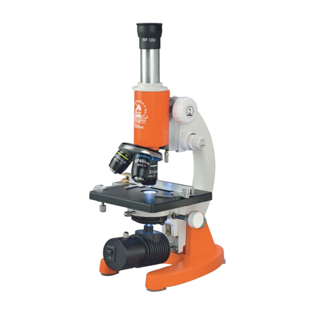
Compound Microscope
A compound microscope lets you see tiny objects on a glass slide. It has two lenses. The objective lens sits near the object and forms a small image. The eyepiece lens enlarges this image for your eyes. It gives higher magnification than simple microscopes and reduces color errors. Doctors, students, and scientists use it to study cells, bacteria, and tissues
Diagram:

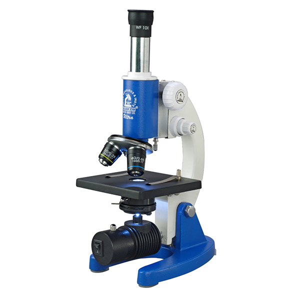
Binocular microscope

A binocular microscope is a type of microscope with two eyepieces. It allows you to look with both eyes at the same time. This makes viewing more comfortable and reduces eye strain. The lenses work like a compound microscope, with an objective lens near the specimen and an eyepiece lens for magnification. It is used to study cells, tissues, and small objects in labs.
Diagram:

Digital Microscope
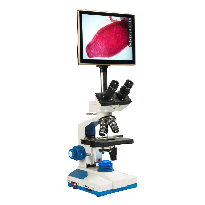
A digital microscope is a microscope that shows the magnified image on a computer or screen. It uses a camera instead of traditional eyepieces. You can capture photos and videos of the specimen. It allows multiple people to view the object at the same time. It is used in schools, labs, and industries to study cells, insects, materials, and small defects in objects.
Diagram:
Stereo Microscope
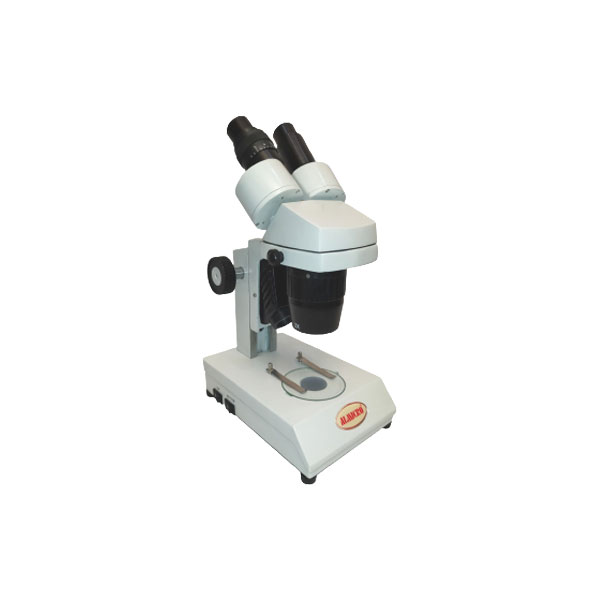
A stereo microscope is a low-power microscope that shows objects in 3D. It has two eyepieces and two separate optical paths. This creates a three-dimensional view of the specimen. It is used to study insects, plants, circuit boards, and small mechanical parts. It does not give very high magnification but helps see shapes and surfaces clearly. It is useful for detailed work and inspection.
Diagram:

5. Fluorescence Microscope
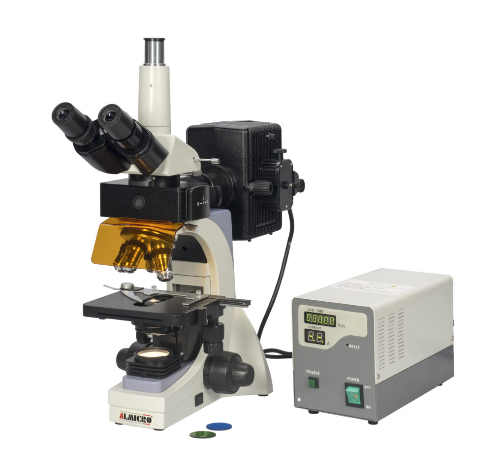
A fluorescence microscope is a microscope that uses special light to make certain parts of a specimen glow. It highlights specific structures like cells, proteins, or tissues. Fluorescent dyes or markers are used to show these parts clearly. Scientists use it to study cells, bacteria, and biological processes. It helps see details that are invisible under normal light.
Diagram:
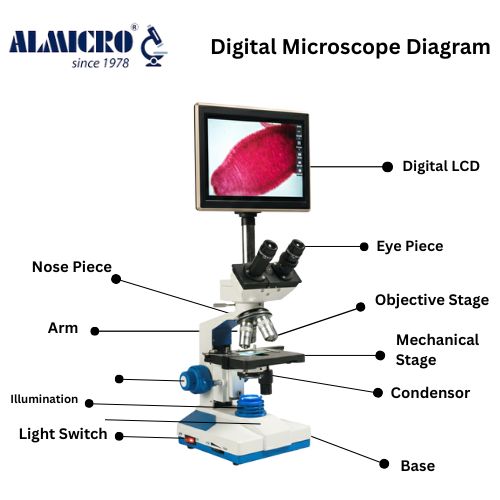
List the main components one by one in Every Microscope. For example:
- Eyepiece – Lens you look through, magnifies the image.
- Objective lenses – Close to specimen, create first magnified image.
- Nosepiece – Holds objective lenses, rotates to change magnification.
- Stage – Flat platform to place the slide, holds specimen.
- Stage clips – Keep the slide in place.
- Light source – Shines light through the specimen to see details.
- Diaphragm – Controls the amount of light passing through the specimen.
- Coarse focus knob – Moves stage or lenses quickly for general focus.
- Fine focus knob – Adjusts focus slowly for clear, sharp image.
- Arm – Connects base to top, used for holding the microscope.
- Base – Supports the microscope and keeps it stable.
- Condenser – Focuses light onto the specimen for better clarity.
History of Microscopy
2.1 Early Developments

The origins of the microscope are generally traced to late sixteenth-century Netherlands, where spectacle-makers began experimenting with combinations of lenses to achieve magnification. Zacharias Janssen and his father Hans Janssen of Middelburg are widely credited with the construction of one of the first compound microscopes around 1590–1595, although historical attribution remains a matter of scholarly debate. Their early instruments, while crude by modern standards, demonstrated that multiple lenses arranged in sequence could produce significantly greater magnification than any single lens alone.
Galileo Galilei, primarily renowned for his telescopic astronomical observations, also constructed a compound microscope around 1609–1610, which he termed the occhiolino (little eye). Although Galileo’s microscopic work was not as influential as his astronomical discoveries, it demonstrated the versatility of lens-based optical instruments. Cornelis Drebbel of the Netherlands independently constructed a compound microscope around the same period, contributing to the growing awareness of microscopy’s scientific potential across Europe.
2.2 Contributions of Antonie van Leeuwenhoek and Robert Hooke
The true scientific potential of the microscope was realised in the seventeenth century through the independent contributions of two pivotal figures: Robert Hooke and Antonie van Leeuwenhoek.
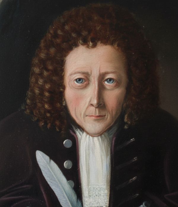
Robert Hooke, an English natural philosopher, published his landmark work Micrographia in 1665 — one of the most significant scientific publications of the seventeenth century. Using a compound microscope of his own construction and improvement, Hooke produced exquisitely detailed illustrations of a wide variety of specimens including cork, fleas, lice, and textiles. Most notably, Hooke’s examination of thin sections of cork revealed a structure of small compartments which he described as cells — the first recorded use of this term in a biological context. Micrographia stimulated widespread public and scientific interest in microscopy throughout Europe.

Antonie van Leeuwenhoek, a Dutch draper and self-taught scientist, achieved an even more remarkable level of optical craftsmanship. Between the 1670s and the early eighteenth century, Leeuwenhoek ground and polished single lenses of extraordinary quality, achieving magnifications of up to approximately 270× — far surpassing any contemporary instrument. Using these simple microscopes, he was the first to observe and describe bacteria (which he termed animalcules), protozoa, spermatozoa, red blood cells, and the capillary circulation of blood in living tissue. His meticulous observations, communicated to the Royal Society of London, established microbiology as a scientific discipline and provided the first empirical glimpses into the invisible world of microorganisms.
2.3 Evolution from Optical to Modern Microscopes
The eighteenth and nineteenth centuries witnessed steady improvements in optical microscopy. Chromatic aberration — the production of colour fringing due to different wavelengths of light refracting at different angles — was substantially reduced through the development of achromatic lenses, pioneered by Chester Moor Hall and later refined by John Dollond in the mid-eighteenth century. In 1830, Joseph Jackson Lister (father of the antiseptic surgeon Joseph Lister) developed a method for combining lenses to eliminate spherical aberration, dramatically improving image sharpness.
Ernst Abbe’s theoretical contributions in the 1870s, developed in collaboration with Carl Zeiss at the Jena optical works, established the mathematical foundations of modern microscope optics. Abbe formulated the diffraction-based resolution limit — now known as the Abbe diffraction limit — which defined the theoretical boundary of resolving power for light-based microscopes as approximately half the wavelength of the illuminating light, or roughly 200 nanometres under visible light conditions. Abbe also developed the apochromatic objective lens, which corrected both chromatic and spherical aberrations to a high degree.
The twentieth century brought revolutionary advances that transcended the physical limits of light. Ernst Ruska and Max Knoll developed the first electron microscope in Germany in 1931–1932, using an electron beam instead of visible light to achieve resolution at the atomic scale. Ruska was awarded the Nobel Prize in Physics in 1986 for this achievement. The scanning tunnelling microscope, developed by Gerd Binnig and Heinrich Rohrer at IBM Zürich in 1981, achieved imaging at the level of individual atoms and earned its inventors the Nobel Prize in Phys
3. Principles of Microscopy
3.1 Magnification
Magnification is the ratio of the apparent size of an object as viewed through the microscope to its actual physical size. In a compound optical microscope, total magnification is the product of the magnification of the objective lens and that of the eyepiece (ocular) lens. For example, a 40× objective combined with a 10× eyepiece yields a total magnification of 400×. Magnification alone does not determine the quality or utility of a microscopic image; without sufficient resolution, increased magnification merely produces a larger but blurred image — a phenomenon referred to as empty magnification.
3.2 Resolution
Resolution is the ability of a microscope to distinguish two closely spaced points as separate entities rather than as a single merged feature. It is the most critical performance parameter of any microscope. The theoretical resolution limit of a light microscope, as formulated by Ernst Abbe, is expressed as: d = λ / (2 × NA), where d is the minimum resolvable distance, λ is the wavelength of the illuminating light, and NA is the numerical aperture of the objective lens. Under optimal conditions using visible light (wavelength approximately 400–700 nm), this limit is approximately 200 nanometres. Electron microscopes, using electron beams with wavelengths measured in picometres, achieve atomic-scale resolution of 0.1–0.2 nanometres or better.
3.3 Contrast
Contrast refers to the difference in intensity or colour between a specimen and its background, and between different features within the specimen. Many biological specimens are inherently transparent and produce little intrinsic contrast in transmitted bright-field illumination. Various techniques have been developed to enhance contrast, including staining with chemical dyes, phase contrast microscopy (developed by Frits Zernike, Nobel Prize 1953), differential interference contrast (DIC) microscopy, dark-field illumination, and fluorescence microscopy. Each technique exploits different physical properties of light and the specimen to reveal structural detail that would otherwise be invisible.
3.4 Illumination Techniques
Proper illumination is essential for high-quality microscopic imaging. Köhler illumination, developed by August Köhler in 1893, is the standard illumination technique used in modern laboratory microscopes. It provides uniform, glare-free illumination across the entire field of view by focusing the image of the light source at the condenser aperture diaphragm rather than directly on the specimen. The illumination sources used in modern microscopes include tungsten-halogen lamps, mercury and xenon arc lamps (for fluorescence applications), light-emitting diodes (LEDs), and laser sources (for confocal and super-resolution systems). For electron microscopes, the electron beam is generated by thermionic emission from a tungsten filament or by field emission from a sharp tungsten tip.
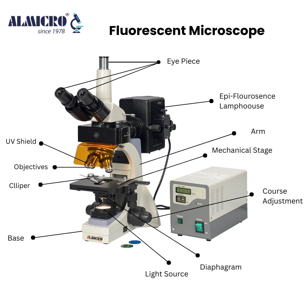
4. Components of a Microscope
4.1 Mechanical Components
The mechanical framework of a compound microscope comprises the base (foot), which provides a stable platform; the arm (limb), which connects the base to the head and body tube; the body tube, which houses the optical components; the nosepiece (revolving turret), which carries the objective lenses; the coarse and fine adjustment knobs, which control the vertical position of the stage or body tube to bring specimens into focus; and the mechanical stage, which holds the microscope slide and permits controlled, reproducible lateral movement in the x and y axes. The quality of the mechanical components, particularly the smoothness and precision of the stage movements and focusing mechanism, significantly affects the ease and accuracy of microscopic observation.
4.2 Optical Components
The optical system of a compound microscope consists of the objective lenses, which are the primary image-forming elements; the eyepiece (ocular) lenses, which provide the secondary magnification viewed by the observer; the condenser, which concentrates and shapes the illuminating light to fill the objective’s numerical aperture; the field diaphragm, which controls the diameter of the illuminated field; and the aperture diaphragm, which controls the angle and intensity of the illuminating cone of light. Modern research microscopes also incorporate a camera port or c-mount adapter for digital imaging, as well as optional beam-splitting modules for simultaneous multi-channel fluorescence detection.
4.3 Illumination System
The illumination system of a modern compound microscope comprises the light source (LED, tungsten-halogen lamp, or arc lamp), collector lenses that gather and direct the light from the source, a field lens, the field diaphragm, the condenser, and the aperture diaphragm. In epi-fluorescence configurations, the illumination path is reflected through the objective lens onto the specimen by a dichroic mirror. Proper alignment and setting of the illumination system according to Köhler principles is essential for optimal imaging performance. LED-based illumination systems have become the dominant technology in modern microscopes due to their long operational lifetime, stability, low heat generation, and narrow spectral output, which is advantageous for fluorescence applications.
5. Working Mechanism
5.1 Image Formation in Optical Microscopes
In a compound optical microscope, the specimen is placed on the stage and illuminated from below by the transmitted light system (for thin, transparent specimens) or from above (for incident, reflected light applications). Light transmitted through or reflected from the specimen enters the objective lens, which focuses the rays to form a real, inverted, magnified intermediate image at a defined plane within the body tube (the intermediate image plane). This intermediate image is then magnified a second time by the eyepiece, which presents a virtual, further-magnified image to the observer’s eye at a comfortable viewing distance (conventionally 250 mm, the near point of the relaxed human eye).
The quality of the final image depends on the aberration correction of both objective and eyepiece lenses, the alignment of the optical axis, the setting of the condenser and diaphragms, and the cleanliness of all optical surfaces. Objective lenses are classified by their degree of aberration correction: achromats correct chromatic aberration for two wavelengths; fluorites (semi-apochromats) offer better correction; and apochromats correct chromatic aberration for three or more wavelengths while also correcting spherical aberration, providing the highest image quality.
5.2 Light Path and Magnification Process
In the standard infinity-corrected optical design used in virtually all modern research microscopes, the objective lens produces a collimated (parallel) beam of light rather than a converging beam directed to a fixed intermediate image plane. A tube lens within the microscope body then focuses this collimated beam to form the intermediate image. This infinity-corrected design allows optical accessories such as fluorescence filter cubes, differential interference contrast (DIC) prisms, and beam splitters to be inserted into the parallel beam path between the objective and tube lens without introducing optical aberrations or shifting the focal plane — a significant practical advantage over older fixed-tube-length designs.
6. Applications of Microscopes
6.1 Medical and Clinical Diagnostics
Microscopy is a cornerstone of clinical pathology and medical diagnosis. Light microscopy of stained tissue sections (histology) is the definitive method for diagnosing the majority of cancers, inflammatory diseases, and structural tissue pathologies. Haematology relies on microscopic examination of blood films (peripheral blood smears) for the identification of abnormal cell types, parasites (such as the malaria-causing Plasmodium species), and haematological malignancies. Clinical microbiology employs both light microscopy (including Gram staining, Ziehl-Neelsen staining for acid-fast bacteria, and direct wet preparations) and fluorescence microscopy for the detection and identification of bacterial, fungal, and parasitic pathogens. Electron microscopy plays a role in the diagnosis of viral infections and ultrastructural pathologies of subcellular organelles in renal biopsy specimens.
6.2 Biological Research
Microscopy has been the primary tool of biological investigation for over three and a half centuries. In cell biology, fluorescence and confocal microscopy enable the real-time imaging of live cells, the tracking of individual molecules, and the three-dimensional reconstruction of subcellular architecture. In developmental biology, time-lapse microscopy documents the dynamic processes of embryonic development. Structural biology employs cryo-electron microscopy (cryo-EM) — a specialised TEM technique involving the rapid freezing of specimens in vitreous ice — to determine the three-dimensional structures of macromolecular complexes, viruses, and membrane proteins at near-atomic resolution, a capability recognised by the Nobel Prize in Chemistry in 2017.
6.3 Material Science
SEM and TEM are indispensable tools in materials science for characterising microstructure, grain boundaries, defects, phase distributions, and elemental composition (via energy-dispersive X-ray spectroscopy, EDS) in metals, ceramics, polymers, composites, and nanomaterials. AFM provides nanometre-scale surface topography and mechanical property data for thin films, coatings, and nanostructured materials. Polarised light microscopy is used for the characterisation of minerals, polymers, and liquid crystals. Confocal Raman microscopy enables chemical mapping of materials at the micrometre scale.
6.4 Industrial and Quality Control
In semiconductor manufacturing, optical and electron microscopes are used for photomask inspection, wafer defect analysis, and the measurement of critical dimensions in integrated circuit fabrication. The pharmaceutical industry employs microscopy for particle size analysis of active pharmaceutical ingredients, the characterisation of crystal polymorphs, and quality control of tablet coatings. The food industry uses microscopy for the detection of adulterants, the assessment of food microstructure, and the identification of foreign material. Forensic science relies on microscopy for the comparison of fibres, hair, soil, pollen, glass fragments, and ballistic evidence.
6.5 Education
Microscopes are fundamental teaching instruments at every level of science education, from primary school onwards. Compound microscopes in school and undergraduate laboratories introduce students to cell theory, microbiology, histology, and materials characterisation. The hands-on operation of microscopes develops practical scientific skills including specimen preparation, optical alignment, quantitative measurement, and critical observation. Digital microscopes with integrated cameras and computer displays enhance group teaching by enabling shared observation and image analysis.
7. Advantages and Limitations
7.1 Advantages
- Optical microscopes are relatively inexpensive, require minimal specimen preparation, and permit the observation of living specimens in real time.
- Electron microscopes provide resolution at the nanometre and sub-nanometre scale, enabling direct visualisation of atomic structures, molecular complexes, and surface topography at a level impossible with light microscopes.
- Fluorescence and confocal microscopes enable highly specific labelling and three-dimensional imaging of defined molecular species within biological specimens.
- Scanning probe microscopes allow measurement of surface properties — including topography, elasticity, electrical conductivity, and magnetic domains — with atomic precision.
- Digital microscopy systems enable remote observation, automated image acquisition, quantitative image analysis, and integration with artificial intelligence for pattern recognition.
7.2 Limitations
- Optical microscopes are fundamentally constrained by the Abbe diffraction limit to a resolution of approximately 200 nm, insufficient for the direct visualisation of individual molecules or atomic structures.
- Electron microscopes require high-vacuum environments and extensive specimen preparation (fixation, dehydration, heavy metal staining or cryogenic preservation), precluding the direct observation of living material under conventional conditions.
- High-performance electron and scanning probe microscopes represent a substantial capital investment, require specialised facilities and technical expertise, and are associated with significant operating costs.
- Sample preparation artefacts are a persistent concern in all forms of microscopy; chemical fixation, staining, or the mechanical scanning of a probe across a surface may alter the specimen from its native state.
- The field of view in high-magnification microscopy is necessarily very small, making comprehensive surveys of large specimens labour-intensive unless automated scanning and image montaging systems are employed.
8. Recent Advances and Innovations
8.1 Super-Resolution Microscopy
A suite of optical super-resolution techniques developed primarily in the 2000s overcomes the Abbe diffraction barrier using photophysical and computational approaches to achieve spatial resolutions of 20–50 nm — more than four-fold below the classical resolution limit — using visible light. The three principal families of super-resolution microscopy are: stimulated emission depletion (STED) microscopy, developed by Stefan Hell; structured illumination microscopy (SIM); and single-molecule localisation microscopy (SMLM), which encompasses photoactivated localisation microscopy (PALM) and stochastic optical reconstruction microscopy (STORM). Stefan Hell, Eric Betzig, and William Moerner shared the Nobel Prize in Chemistry in 2014 for the development of super-resolved fluorescence microscopy.
8.2 Cryo-Electron Microscopy
Cryo-electron microscopy (cryo-EM) has undergone a transformational advance in the 2010s, driven by the development of direct electron detection cameras with improved sensitivity and the application of sophisticated computational algorithms for single-particle image reconstruction. These advances — sometimes referred to as the resolution revolution in cryo-EM — have enabled the routine determination of protein and macromolecular complex structures at resolutions of 2–4 ångströms, approaching and in some cases matching the resolution of X-ray crystallography, without the requirement for crystallisation. Cryo-EM is now a primary structural biology technique of immense pharmaceutical relevance.
8.3 Digital Microscopy and AI Integration
The integration of high-resolution digital cameras, motorised stages, and sophisticated image analysis software has transformed light microscopy into an automated, high-throughput imaging platform. High-content screening systems perform automated acquisition and analysis of fluorescence images from thousands of wells of cultured cells, enabling genome-wide genetic screens and drug discovery assays at a scale impossible by manual microscopy. Artificial intelligence — particularly deep learning convolutional neural networks — is increasingly applied to the automated classification of histological slides for cancer diagnosis, the segmentation of cellular structures, the enhancement of image quality, and the prediction of molecular features from morphological images (computational pathology). Whole-slide imaging (WSI) systems digitise entire histological glass slides at high resolution, facilitating remote pathology consultation and digital archiving.
9. Maintenance and Calibration
9.1 Cleaning Procedures
The optical performance of a microscope is critically dependent on the cleanliness of all optical surfaces. Dust, fingerprints, immersion oil residues, and biological specimens can degrade image quality and, if left unattended, permanently damage coated lens surfaces. External optical surfaces (eyepieces, objectives, condenser) should be cleaned using lint-free optical tissues moistened with appropriate solvents (lens cleaning solution, isopropanol, or diethyl ether–ethanol mixture). Immersion oil must be removed from oil-immersion objectives after each use. Internal optical components should not be cleaned by non-specialist personnel; if internal contamination is suspected, the instrument should be returned to the manufacturer or a qualified service engineer.
9.2 Calibration Standards
Accurate quantitative microscopy requires regular calibration of the imaging system. Lateral (x-y) magnification calibration is performed using certified stage micrometers — glass slides engraved with a precise scale — imaged at each objective magnification. Axial (z-axis) calibration for confocal and other three-dimensional imaging systems requires specialised reference targets. Resolution is routinely assessed using fluorescent bead preparations of defined size (typically 100–200 nm diameter fluorescent polystyrene microspheres) to measure the point spread function of the instrument. In clinical laboratories, calibration procedures for quantitative measurements (e.g., cell counting, particle sizing) are subject to accreditation standards and must be documented and traceable to national measurement standards.
9.3 Common Operational Issues
Frequently encountered operational problems include: incorrect Köhler illumination alignment resulting in uneven background illumination; accumulation of immersion oil on dry objectives causing image haze; mould growth on optical elements in humid environments; mechanical drift of the focus and stage in older instruments; image vibration from building vibration sources (requiring pneumatic or active anti-vibration tables for high-magnification work); lamp instability in mercury and xenon arc sources; and photobleaching of fluorescent specimens during extended observation with intense illumination.
10. Safety Considerations
10.1 Electrical Safety
All electrically powered microscopes present potential electrical hazards if improperly maintained or operated. High-voltage power supplies are required for mercury and xenon arc lamps used in fluorescence microscopy; these lamps must be allowed to cool fully before handling and must be replaced by trained personnel, as they operate under pressure and may explode if mishandled. Laser sources used in confocal and super-resolution systems are classified as Class 3B or Class 4 hazards and require appropriate laser safety training, interlock systems, and personal protective equipment. Electron microscopes involve high accelerating voltages (up to 300–400 kV in research TEMs) and require strict adherence to electrical safety protocols.
10.2 Biological Hazards
The preparation and observation of biological specimens under the microscope may involve exposure to infectious agents, chemical fixatives, and staining reagents that present biological and chemical hazards. Specimens potentially containing pathogenic microorganisms, bloodborne pathogens, or prion-infected material must be handled in accordance with applicable biosafety level (BSL) regulations and institutional biosafety committee guidelines. Chemical fixatives such as formaldehyde and glutaraldehyde are toxic and, in the case of formaldehyde, known carcinogens; their use requires adequate fume hood ventilation and appropriate personal protective equipment. Heavy metal stains used in electron microscopy (osmium tetroxide, uranyl acetate, lead citrate) are highly toxic and require specialised handling, storage, and disposal procedures.
10.3 Proper Handling
Microscopes are precision instruments that require careful handling. They should always be carried with two hands — one supporting the base and one holding the arm — and should be stored covered to prevent dust accumulation. Glass slides should be handled with gloves where infection risk is present, and broken slides disposed of in appropriate sharps containers. Objective lenses should never be touched with bare fingers. The coarse focus knob should never be turned while observing with oil-immersion objectives to avoid crushing slides and damaging expensive objective lenses. All personnel should
Fun Facts About Microscopes
- Anton van Leeuwenhoek called bacteria “animalcules” in his letters.
- Robert Hooke named cells after the rooms in a monastery.
- Early microscopes could only magnify objects 20–30 times.
- Some of Leeuwenhoek’s microscopes were less than 3 cm long.
- The word “microscope” comes from Greek words meaning “small” and “to look.”
- Modern electron microscopes can magnify objects over 10 million times.
- Scientists use microscopes to study art, like analyzing pigments in old paintings.
- Microscopes are used in forensic labs to detect tiny evidence in crimes.
- Without microscopes, bacteria, viruses, and tiny cells would remain invisible to humans.
Frequently Asked Questions
A microscope is a scientific instrument used to see and study objects that are too small to be seen with the naked eye. It works by using lenses or other technologies to magnify tiny objects, allowing us to observe details such as cells, bacteria, fibers, and fine surface structures clearly.
A microscope is needed because the human eye cannot see very small objects like cells, bacteria, and fine surface details. It helps scientists and doctors study things that are invisible to the naked eye.
The first compound microscope was invented by Zacharias Janssen in the late 16th century. His invention used two lenses to achieve greater magnification.
Most microscopes include an eyepiece, objective lenses, stage, light source, focus knobs, arm, base, and condenser. These parts work together to produce a clear magnified image.
Robert Hooke discovered and named “cells” after observing cork under a microscope, marking an important milestone in the history of biology.
Anton van Leeuwenhoek was the first to observe bacteria and microorganisms, revealing a hidden world of microscopic life and advancing biological science.
Modern microscopes offer much higher magnification, better clarity, digital displays, and even atomic-level imaging, unlike early microscopes which were simple and low-powered.
Microscopes are used in medicine, education, research, industry, forensics, and material testing to examine tiny details and solve real-world problems.