1. What is a Phase Contrast Microscope?
A phase contrast microscope is a special type of optical microscope designed to observe transparent and colorless specimens without staining. It works by enhancing the contrast between different structures in the specimen that have slight variations in refractive index.
Unlike a traditional bright-field microscope, which struggles to show details in unstained living cells, the phase contrast microscope converts invisible phase shifts in light into visible brightness changes — allowing scientists to see living cells, organelles, and microorganisms clearly in their natural state.
2. Phase Contrast Microscope Diagram
Below is a simple phase contrast microscope diagram showing its key components such as the light source, condenser annulus, specimen, objective lens, and phase plate.
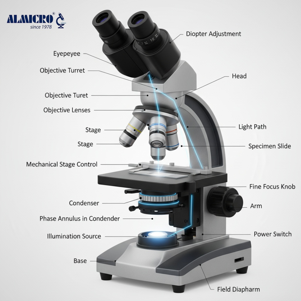
(Insert a labeled image here with “Phase Contrast Microscope Diagram” as the alt text.)
Each part plays a vital role:
-
Condenser annulus: Shapes the light cone that passes through the specimen.
-
Specimen stage: Holds the slide in place for observation.
-
Objective lens: Collects the light and magnifies the image.
-
Phase plate: Introduces a controlled phase shift to enhance contrast.
This phase contrast microscope diagram helps visualize how the optical path changes to produce detailed images.
3. History and Invention of Phase Contrast Microscopy
The credit for inventing the phase contrast microscope goes to Dutch physicist Frits Zernike in 1934. Zernike’s phase contrast technique enabled investigators to see living cells easily precisely because it avoided the use of staining, which was a significant advance for biological research.

For this achievement, he was awarded the Nobel Prize in Physics in 1953. Prior to the invention of the phase contrast microscope, live transparent samples were virtually impossible to observe – staining them would quickly kill them or ultimately alter them. The phase contrast technique revolutionized microscopy to better study cell division, motility, and cell´s internal structures in real-time.
4. Phase Contrast Microscope Principle (Explained Simply)
The phase contrast microscope principle is based on the fact that light waves passing through transparent specimens experience small phase shifts — these are invisible to the human eye. The microscope converts these phase differences into brightness or intensity variations.
Simply put:
-
When light passes through thicker or denser areas of the sample, its speed changes slightly.
-
These tiny changes cause phase shifts.
-
The phase plate inside the microscope transforms these shifts into contrast differences, creating a visible image.
This process lets you see living cells and transparent structures without using stains or dyes.
5. Working of a Phase Contrast Microscope (Step-by-Step)
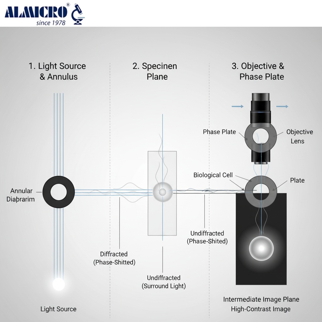
Here’s how the phase contrast microscope works step by step:
-
Light Source: Emits light that passes through the condenser annulus.
-
Condenser Annulus: Directs a hollow cone of light toward the specimen.
-
Specimen Interaction: Different parts of the specimen change the phase of light differently.
-
Objective Lens: Collects both direct (undeviated) and diffracted light.
-
Phase Plate: Located in the objective; it introduces a specific phase shift.
-
Interference: The direct and diffracted light interfere, producing bright and dark regions.
-
Image Formation: The final image appears with high contrast, revealing internal cell details.
This simple yet powerful working principle makes the phase contrast microscope essential in every biology and medical lab.
6. Purpose of Phase Contrast Microscopy
The phase contrast microscope has countless applications in research, medicine, and education. Some of the most common uses include:
-
Cell Biology: Observing live, unstained cells and organelles.
-
Microbiology: Viewing bacteria, protozoa, and single-cell organisms.
-
Hematology: Studying blood cells without stains.
-
Embryology: Monitoring early embryo development.
-
Tissue Culture: Examining cells in petri dishes without disturbing growth.
-
Educational Labs: Demonstrating live cell structures to students.
Because it allows visualization without stains or dyes, the phase contrast microscope is ideal for studying living organisms in their natural environment.
7. Advantages and Limitations
Advantages
-
Enables observation of living, unstained cells.
-
Produces high-contrast images of transparent samples.
-
Simple and quick setup for routine lab use.
-
Non-destructive — specimens remain alive and unaltered.
Limitations
-
Produces halo artifacts around the image edges.
-
Not ideal for thick or opaque specimens.
-
Phase objectives are more expensive than standard ones.
-
Image interpretation requires experience.
Despite these drawbacks, its benefits far outweigh the limitations in biological and clinical research.
8. Principle of Phase Contrast Microscope (Detailed Version)
The concept behind a phase contrast microscope uses differences in the optical path length and wave interference.
When light is transmitted through a transparent specimen, certain parts of the specimen will slow the light more than others, causing phase differences. These phase differences are usually on the order of 1/4 of a wavelength, which cannot be perceived by the naked eye.
The phase plate in the objective lens introduces an additional wave advance or retardation to part of the light waves equal to another 1/4 of a wavelength. When the two light waves, the diffracted (specimen) and undiffracted (background), are back together, they will add constructively or destructively when they interfere, and this results in bright and dark contrasts in the final image.
This more complex notion of optical interference is the basis for the effectiveness of the phase contrast microscope principle for imaging biological specimens.
9. Summary – Why It’s a Must-Have in Modern Labs
In modern laboratories, the phase contrast microscope is indispensable. Its ability to display clear, detailed images of live, unstained specimens has made it a cornerstone of life science research.
From tracking cellular behavior to observing microorganisms in real time, this microscope helps researchers see life as it truly is — without artificial stains or chemicals. Every educational, medical, and research lab benefits from having one. Looking for more microscope types? Click Here 👈
10. FAQs About Phase Contrast Microscope
Q1. Who invented the phase contrast microscope?
Frits Zernike invented it in 1934 and received the Nobel Prize in 1953.
Q2. What is the principle of a phase contrast microscope?
It converts phase shifts in light waves passing through a specimen into brightness differences, enhancing contrast.
Q3. Why is the phase contrast microscope used?
It’s used to study living, transparent cells without staining.
Q4. What are the limitations of phase contrast microscopy?
Halo formation and unsuitability for thick samples are common limitations.
Q5. What type of specimens are best observed using this microscope?
Transparent, colorless, or living biological samples such as cells, protozoa, and bacteria.