
Phase Contrast Microscope vs Fluorescence Microscope: Key Differences Explained
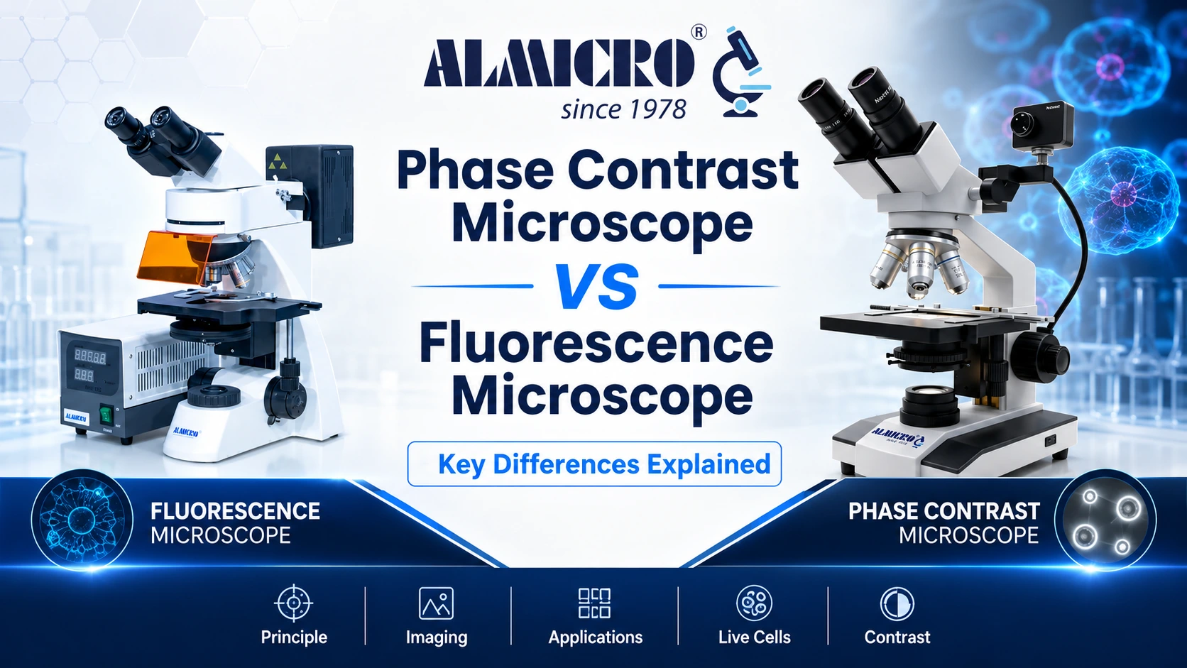
A phase contrast microscope converts phase shifts in light into brightness differences, allowing unstained living cells to be observed with minimal sample preparation. A fluorescence microscope uses fluorophores and excitation light to reveal specific labelled structures against a dark background. The two techniques solve different problems. Choosing the wrong one can mean paying for capabilities a laboratory will rarely use or missing the molecular specificity a project actually requires.
Phase Contrast vs Fluorescence Microscopy: Quick Comparison
| Parameter | Phase Contrast Microscope | Fluorescence Microscope |
|---|---|---|
| Working principle | Converts phase shifts into visible intensity differences | Excites fluorophores and detects their emitted light |
| Sample preparation | Minimal; generally no staining required | Fluorescent staining, tagging, or naturally fluorescent material required |
| Light source | Halogen or LED white light | LED, mercury, or metal-halide excitation source |
| Key optics | Phase annulus and matching phase plate | Excitation filter, dichroic mirror, and emission filter |
| Specificity | Shows general cell morphology and dynamics | Can reveal specific labelled proteins, organelles, or molecular targets |
| Live-cell imaging | Excellent and non-destructive under suitable conditions | Possible, but limited by phototoxicity and photobleaching |
| Typical cost | Lower | Higher |
| Common uses | Cell-culture checks, motility studies, and contamination screening | Immunofluorescence, apoptosis studies, and molecular localisation |
What Is a Phase Contrast Microscope?
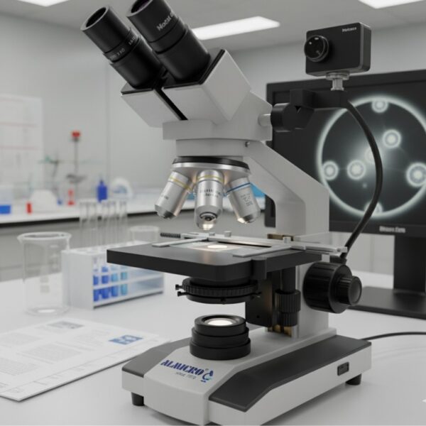
Phase Contrast Microscope
A phase contrast microscope is a compound light microscope modified to reveal detail in transparent, unstained specimens. Instead of relying mainly on colour or absorption, it uses small changes in the phase of light as the light passes through structures with different thicknesses or refractive indices.
How Phase Contrast Works
Light passing through a cell is delayed slightly compared with light passing through the surrounding medium. This phase shift does not create visible contrast by itself. A phase annulus in the condenser and a matching phase plate in the objective convert the phase difference into an intensity difference. As a result, cell boundaries and many internal structures become easier to observe.
The technique was developed by Frits Zernike, who received the Nobel Prize in Physics in 1953 for the phase contrast method. It remains one of the standard approaches for observing living cells without staining them first.
Key Components
A phase contrast setup requires a condenser fitted with a phase annulus, phase objectives containing matching phase plates, and usually a centring telescope or alignment aid. Correct alignment is essential because contrast and image quality decline when the condenser annulus and objective phase ring do not match properly.
Where Phase Contrast Is Used
Phase contrast is widely used in cell-culture laboratories for confluency checks, contamination screening, sperm-motility analysis, and real-time observation of bacteria or protozoa. It is especially useful when a specimen needs to remain alive and unstained. Inverted tissue-culture microscopes therefore commonly include phase contrast for routine observation of cells growing in flasks, dishes, and multi-well plates.
What Is a Fluorescence Microscope?
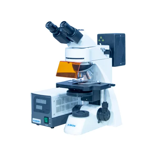
Fluorescence Microscope
A fluorescence microscope is designed to reveal the locations of fluorescent labels or naturally fluorescent molecules within a specimen. Unlike phase contrast, which displays general morphology, the fluorescence channel can highlight selected cells, proteins, organelles, microorganisms, or other targets.
Read Full Fluorescence Guide →
How Fluorescence Microscopy Works
A fluorophore absorbs light within a particular excitation range and emits light at a longer wavelength. The separation between the excitation and emission peaks is called the Stokes shift. An excitation filter selects the incoming wavelength, a dichroic mirror directs it toward the specimen, and an emission filter blocks most unwanted light while allowing the selected fluorescence signal to reach the eyepiece or camera.
Key Components
A fluorescence microscope requires a suitable illumination source, filter sets matched to the fluorophores, and objectives capable of collecting the comparatively weak emitted light. Mercury and metal-halide lamps have traditionally been used, while LED excitation is increasingly common because it offers stable output, lower heat, and longer service life.
Where Fluorescence Is Used
Fluorescence microscopy is used for immunofluorescence, fluorescence in situ hybridisation (FISH), live-cell tracking, cancer research, and other work requiring the localisation of specific markers. It is also used in selected diagnostic and environmental applications, including auramine-based screening for acid-fast bacilli and fluorescence methods for detecting target organisms in water samples.
Phase Contrast vs Fluorescence Microscope: Which One Do You Need?
The right choice depends on the observation task, not on which system sounds more advanced.
- Routine cell culture and morphology: Phase contrast normally provides the required information at a lower purchase and running cost.
- Specific molecular targets or multicolour labelling: Of these two techniques, fluorescence is the appropriate choice because phase contrast cannot distinguish one labelled protein or molecular target from another.
- Both requirements in one laboratory: A trinocular microscope configured with phase contrast and fluorescence attachments can provide a practical combined workflow without requiring two separate microscope stands.
As a laboratory equipment manufacturer based in Ambala Cantt, Haryana, operating since 1978, ALMICRO supplies phase contrast and fluorescence configurations across its microscope range. Laboratories should select a configuration according to specimen type, required targets, imaging frequency, documentation needs, and budget rather than availability alone.
Advantages and Limitations of Each Technique
Phase Contrast Advantages and Disadvantages
Phase contrast is highly practical for live-cell observation. It generally requires no staining, helps cells remain viable during routine examination, has relatively low running costs, and is straightforward to maintain once the optical components are aligned.
Its limitations are visible in the image. Bright halos can appear around specimen edges, while shade-off can reduce contrast across broad or thick structures. Dense specimens may perform poorly, and the method does not provide molecular specificity. It also requires matched phase objectives and a compatible condenser.
Fluorescence Advantages and Disadvantages
Fluorescence provides high specificity. It can reveal selected molecular targets, support multiple labels in different colours, and produce a strong signal against a dark background when the fluorophores, filters, and imaging settings are properly matched.
The disadvantages include photobleaching, in which fluorescence fades during exposure, and phototoxicity, which can damage living cells. Autofluorescence and nonspecific labelling can interfere with the target signal. Fluorescence systems also have higher equipment and maintenance costs, including illumination, filters, cameras, and alignment requirements.
Can You Combine Phase Contrast and Fluorescence?
Yes. Inverted research microscopes commonly combine a phase contrast condenser with fluorescence illumination and filter sets on the same stand.
This arrangement supports a practical workflow. The operator can use phase contrast to locate, focus, and assess the cells before switching to a fluorescence channel to image a labelled target. A trinocular microscope body with both capabilities can complete this workflow without moving the specimen between two separate instruments.
Frequently Asked Questions
What Is Another Name for a Phase Contrast Microscope?
It is sometimes called a Zernike phase contrast microscope after Frits Zernike, who developed the technique. It may also be described as a phase contrast light microscope or simply as phase microscopy.
What Are the Disadvantages of Using a Phase Contrast Microscope?
The main limitations include halo artefacts around specimen edges, shade-off in broad or thick regions, reduced performance with dense specimens, and a lack of molecular specificity. The technique also requires matched phase objectives and a compatible phase condenser that must be aligned correctly.
When Would You Use a Phase Contrast Microscope?
Use phase contrast when specimens are transparent, unstained, and preferably living. Common applications include cell-culture confluency checks, contamination screening, sperm-motility analysis, and observation of bacteria or protozoa when staining could kill or alter the sample.
What Is the Difference Between a Phase Contrast Microscope and a Light Microscope?
A phase contrast microscope is a specialised type of light microscope equipped with a phase annulus and matching phase plate. A standard bright-field microscope relies mainly on absorption and scattering contrast, so transparent unstained cells can be difficult to see. Phase contrast converts otherwise invisible phase differences into visible intensity differences.