

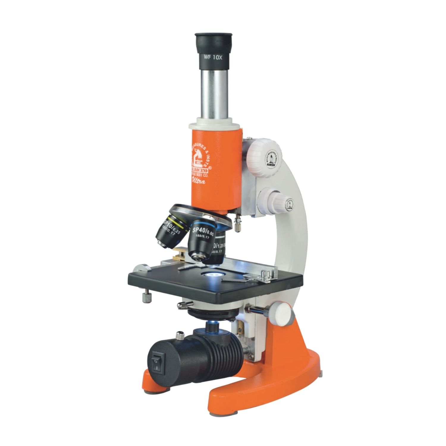
 Definition of a Microscope
A microscope is one of those inventions that completely changed the way we see the world. It gives us a peek into things our eyes can never catch on their own. Put simply, a microscope is a tool that magnifies objects so tiny that they’re invisible to the naked eye. With its help, we can zoom in on the smallest details—like the structure of living cells, bacteria swimming around, delicate tissues, or even the fine grains of metals, fibers, and countless other materials. It’s like unlocking a hidden universe that has always been around us, just waiting to be seen.
Types of Microscope
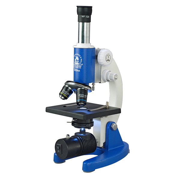 1. Compound Microscope
 The compound microscope is probably the one most people picture first. It’s a light microscope that uses two or more lenses to magnify objects, making it a staple in classrooms, laboratories, and research centers.
“Compound” refers to its two-lens system—the objective lens and the eyepiece—working together to create a highly magnified image. Depending on the lens setup, it can enlarge objects anywhere from about 40x up to 1000x or more. Light passes through a specimen placed on a glass slide, then through the lenses, producing a sharp and enlarged image.
Main parts include:
-
Eyepiece (ocular lens), usually around 10x magnification
-
Objective lenses with varying magnifications (4x, 10x, 40x, 100x)
-
Stage, where the slide is placed
-
Light source or mirror to illuminate the sample
-
Coarse and fine adjustment knobs for focusing
Common uses:
-
Studying cells and tissues in biology
-
Viewing microorganisms such as bacteria and protozoa
-
Examining blood smears and tissue samples in medical labs
-
Teaching students about microscopic structures in education
-
Supporting research into organisms or structures invisible to the naked eye
2. Educational Microscope
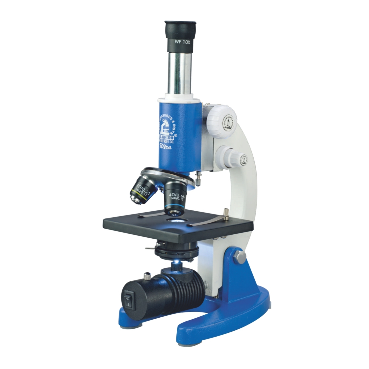
An educational microscope is built with teaching in mind. Found in schools, colleges, and universities, it’s designed to be simple, sturdy, and easy for students to use.
Major Components:
-
Usually a compound light microscope
-
Magnification ranges from 40x to 1000x
-
Basic components like eyepiece, objectives, stage, light source, and focus knobs
-
Affordable and easier to handle than advanced research microscopes
Uses in education:
-
Viewing plant and animal cells
-
Studying bacteria, fungi, protozoa, and algae
-
Bringing life sciences to life through practical observation
-
Encouraging curiosity and hands-on learning in students
Put simply, an educational microscope is a student-friendly tool that makes science tangible and engaging by showing the hidden microscopic world.
3. Stereo Microscope

Also known as a dissecting microscope, a stereo microscope provides a 3D view of the specimen. Unlike compound microscopes, which excel with thin, transparent samples, stereo microscopes are ideal for examining larger, opaque objects at lower magnifications.
Features:
-
Two separate eyepieces and objectives for a true 3D image
-
Low magnification, typically between 10x and 80x
-
Built-in light sources from above and below for different viewing angles
Uses:
-
Biology labs: insects, plant parts, crystals, and rocks
-
Forensic science: examining fibers, hair, or small evidence samples
-
Industry: inspecting electronic parts or machine components
-
Education: teaching surface structures of specimens
-
Hobbies: coin collecting, stamp study, or looking at minerals
In short, stereo microscopes are best when you want a three-dimensional view of solid surfaces and fine details.
4. Medical Microscope

Medical microscopes are designed specifically for hospitals, clinics, and labs. Their role is clear: help detect and study diseases.
Major Components:
-
Often compound light microscopes or advanced digital/electron versions
-
Magnifications ranging from 40x to 2000x or more
-
Built with strong light sources and precision lenses
-
Frequently fitted with digital cameras for documentation
Medical applications:
-
Pathology: analyzing biopsy tissues for cancer or infections
-
Hematology: studying blood cells for anemia, leukemia, or malaria
-
Microbiology: detecting bacteria, fungi, and parasites
-
Cytology: checking body fluids and cells for abnormalities
-
Surgery: advanced surgical microscopes used in neurosurgery, ophthalmology, and dentistry
In medicine, these microscopes aren’t optional. They’re life-saving tools that allow professionals to see what causes disease and guide treatments with accuracy.
5. Phase Contrast Microscope
A phase contrast microscope is designed for one special task: making transparent specimens visible without staining them. This includes living cells, microorganisms, and thin tissues that would normally appear invisible under standard light.

It works by converting differences in light waves that pass through the specimen into variations in brightness and contrast, producing a clear image.
Major Components:
-
Special condenser and phase plates to enhance contrast
-
Allows viewing of live specimens in their natural state
-
Magnification range similar to a compound microscope (40x–1000x)
-
Popular in cell biology, microbiology, and medical labs
Uses:
-
Studying live cells, including their division and movement
-
Identifying microorganisms without the need for staining
-
Medical research on blood cells, sperm, and tissues
-
Observing cell cultures in laboratories
-
Teaching cell biology using living examples
A phase contrast microscope is indispensable when the goal is to study living, unstained cells in detail.
Uses of a Phase Contrast Microscope
Phase contrast microscopes are especially valuable when you need to study living, transparent specimens without staining them. Researchers and students often turn to this tool for tasks like:
-
Watching living cells in action, including their division, movement, and internal activity
-
Identifying microorganisms such as bacteria or protozoa in their natural state
-
Examining blood cells, sperm cells, or tissues for medical research without altering them
-
Monitoring cell growth in tissue culture labs
-
Teaching students about cell biology by showing them live specimens rather than static slides
In short, this type of microscope makes invisible, unstained samples visible. That’s why it has become a staple in biology, medicine, and laboratory research.
6.What is a Binocular Microscope?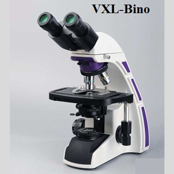
A binocular microscope is an optical microscope designed with two eyepieces, allowing the user to view specimens with both eyes simultaneously. This dual-eyepiece design provides a more comfortable viewing experience and reduces eye strain, especially during long observation sessions. Unlike advanced microscopes that use lasers or digital reconstruction, a binocular microscope relies on visible light and glass lenses to magnify specimens clearly.
It is commonly used for routine laboratory work, education, and medical examination where clear, detailed viewing is required.
Major Components include:
-
Two eyepieces for binocular (dual-eye) viewing
-
Objective lenses with different magnification levels
-
A built-in illumination system (LED or halogen light)
-
A mechanical stage to hold and move the specimen slide
-
Coarse and fine focusing knobs for precise image adjustment
-
A sturdy body designed for stable and long-term use

Key Features:
-
Comfortable viewing with reduced eye fatigue
-
Clear and sharp magnification of biological samples
-
Easy focusing and smooth specimen movement
-
Suitable for prolonged laboratory or classroom use
-
Reliable performance for routine microscopic analysis
Where it’s used:
-
Educational institutions, for teaching biology and life sciences
-
Medical laboratories, for examining blood, tissues, and samples
-
Research labs, for routine biological and chemical analysis
-
Clinical diagnostics, for preliminary disease investigation
-
Industrial labs, for quality inspection and testing
7. What is a Light Microscope?

The light microscope, often called an optical microscope, is probably the most familiar of all. It uses visible light and lenses to magnify specimens that are too small to see with the naked eye, like cells, tissues, or microorganisms. Because it’s simple, affordable, and effective, you’ll find it in schools, universities, labs, and hospitals around the world.
Major Components:
-
Relies on visible light for illumination
-
Uses one or more glass lenses (eyepiece and objectives)
-
Offers magnification from about 40x up to 1000x
-
Works with both live and stained specimens
-
Portable and accessible for routine use
Common applications:
-
Education: helping students view cells, bacteria, and microscopic structures
-
Medicine: assisting doctors and lab technicians in studying blood, tissues, and microbes
-
Research: examining cell division, structure, and microbial activity
-
Industry: checking fibers, electronics, and small materials for quality control
-
Forensics: analyzing hairs, fibers, and other trace evidence
Simply put, the light microscope is the foundation of biological study and a workhorse in medical and scientific settings.
8. What is a Polarizing Microscope?

A polarizing microscope is a specialized type of light microscope designed to work with polarized light. It’s especially useful for studying materials like minerals, crystals, and fibers that interact uniquely with polarized light.
It includes two main filters: the polarizer, placed before the specimen, and the analyzer, positioned after it. Together, they reveal properties of the material that would otherwise remain hidden.
Major Components:
-
Uses polarized rather than standard light
-
Provides structural and compositional details of crystals and minerals
-
Detects birefringence, where materials split light into two separate rays
-
Offers magnification levels similar to a compound microscope (40x–1000x)
Typical uses:
-
Geology and mineralogy, for identifying rocks and minerals
-
Chemistry, for studying salts, crystals, and compounds
-
Forensics, for analyzing fibers, hair, glass, and synthetic materials
-
Material science, for testing plastics, ceramics, and polymers
-
Biology, in some cases, for structures that exhibit birefringence, such as starch grains or muscle fibers
In short, the polarizing microscope is indispensable for fields like geology, chemistry, and forensic science, where understanding material properties is critical.
9. What is a Simple Microscope?

The simple microscope is the most basic version, relying on a single convex lens to magnify an object. It functions much like a magnifying glass, offering a closer look at small details that aren’t visible to the unaided eye.
Historically, it was the very first type of microscope, famously used by Anton van Leeuwenhoek in the 17th century to observe bacteria and tiny organisms no one had ever seen before.
While its magnification is low compared to modern instruments—typically 10x to 20x, sometimes up to 50x—it laid the foundation for modern microscopy and remains useful in certain contexts.
Major Components of a Simple Microscope
-
Uses a single convex lens
-
Low magnification, usually around 10x–20x, sometimes up to 50x
-
Small, lightweight, and easy to carry
-
Less powerful compared to a compound microscope
Uses of a Simple Microscope
-
Education: often used in classrooms for basic magnification demos
-
Biology: viewing small specimens such as algae, fungi, or simple cells in limited detail
-
Jewelry & Watchmaking: jewelers use it to inspect stones and watch parts
-
Forensics: handy for a quick look at fibers or small traces of evidence
-
Everyday tasks: works like a magnifying glass for reading fine print or examining tiny objects
In short, a simple microscope is the earliest form of magnification. It’s a single-lens tool suited for low-power observation.
10. What is a Digital Microscope?


A digital microscope is a modern upgrade. Instead of peering through an eyepiece, you see the magnified specimen directly on a computer, tablet, or monitor. It pairs traditional optical magnification with digital imaging, which makes it easy to capture, store, and even share pictures or videos.
Major Components:
-
Built-in digital camera, sometimes alongside eyepieces
-
Connects via USB, HDMI, or Wi-Fi
-
Magnification usually ranges from 20x up to 1000x or more
-
Can record images and video for study or teaching
-
Easy to use in classrooms, labs, and industry
Common uses:
-
Education: projecting specimens onto a screen so a whole class can see
-
Medical labs: storing and analyzing images of cells, tissues, or blood
-
Research: documenting findings and analyzing samples
-
Industry: checking textiles, electronics, or machine parts for quality control
-
Forensics: recording microscopic evidence for investigation
In short, a digital microscope blends optics with digital tools, making it far easier to share and document microscopic details across medicine, science, and industry.
11. What is an Inverted Microscope?

An inverted microscope works the opposite way of a traditional compound microscope. Here, the objectives sit below the stage, while the light source and condenser are placed above. This setup makes it possible to view specimens in larger containers, like flasks or petri dishes, without disturbing them.
Major Components:
-
Objectives positioned under the stage, with light shining from above
-
Designed for viewing living cells in liquid culture
-
Handles larger specimens that won’t fit on a slide
-
Offers magnification similar to a compound microscope, around 40x–1000x
Where it’s used:
-
Cell culture studies, especially when monitoring live cells
-
Medical research for studying tissues, blood, or microorganisms in natural conditions
-
Embryology, such as tracking fertilization in IVF labs
-
Pharmaceuticals, for drug testing on living cells
-
Material science, where larger samples like metals or alloys need examination
In short, the inverted microscope is ideal when you need to observe living or larger specimens without slicing them into slides.
Microscope Labeled Parts

Uses of a Microscope
1. Microscope in the Medical Field
In medicine, the microscope isn’t a luxury—it’s a necessity. Modern diagnosis would be almost impossible without it. The pathogens and fine structures that hide from the naked eye are laid bare under its lens. Fields like pathology, microbiology, hematology, and clinical research wouldn’t function the same way without this instrument.
Here’s how doctors and lab teams rely on microscopes every day:
-
Detecting bacteria, fungi, and viruses responsible for infections
-
Checking blood cells for diseases like anemia, malaria, or leukemia
-
Examining tissue biopsies to confirm cancers and other conditions
-
Analyzing body fluids, such as urine or sputum, for irregularities or pathogens
-
Supporting surgery with specialized microscopes that magnify delicate structures like nerves, blood vessels, or eye tissue
In Care Services, a microscope isn’t just another piece of lab equipment. It’s a tool that enables early detection, supports precise diagnoses, and guides life-saving treatments. When accuracy and speed are critical, this instrument delivers both.
2. Microscope in Education
In classrooms, the microscope turns abstract lessons into something real. A textbook might explain the structure of a cell, but when a student peers into a microscope and actually sees it, that’s a different experience altogether. That first look often sparks curiosity—and sometimes a lifelong interest in science.
Students use microscopes to:
-
View plant and animal cells and understand how they’re built
-
Explore microorganisms like bacteria, protozoa, and algae
-
Reinforce theories through hands-on lab experiments
-
Build critical thinking skills by examining real specimens
-
Prepare for advanced study with practical work in biology, zoology, and microbiology
Microscopes in education don’t just help students learn facts. They give them a direct view of life’s building blocks, making science far more engaging and memorable.
3. Microscope in Scientific Research
Research labs treat microscopes as more than instruments—they’re the starting point of discovery. In biology, chemistry, physics, and beyond, microscopes give scientists access to worlds at the microscopic and nanoscopic level, producing the kind of detail that no other tool can.
Researchers use them to:
-
Track how cells grow, divide, and communicate with one another
-
Study bacteria, fungi, and viruses while working on new medicines and vaccines
-
Examine DNA, proteins, and molecular structures up close
-
Analyze metals, crystals, and nanomaterials in material science
-
Monitor microorganisms in soil, air, and water for environmental studies
-
Look at fibers, dust, and trace residues for forensic or industrial work
In short, research without microscopes would stall. They expand human knowledge, drive new technologies, and even help address health and environmental problems.
4. Microscope in Forensic Science
Forensics is another field where microscopes play an outsized role. Think of them as the quiet partner in an investigation. Crime scenes often leave behind evidence so small—fibers, hair strands, dust—that it’s invisible without magnification. Under a microscope, though, those tiny traces can tell a bigger story.
Forensic specialists depend on microscopes to:
-
Compare hairs, threads, or fibers to link suspects or victims to locations
-
Examine fingerprints or even skin cells left behind
-
Analyze blood, semen, and other biological material for DNA testing
-
Study residues like gunshot particles, fragments of glass, or bits of soil to reconstruct events
-
Authenticate documents by checking ink, handwriting, and paper fibers under high magnification
In forensic science, the microscope isn’t only about science—it’s about justice. By bringing hidden evidence into focus, it helps build cases that hold up in court.
5. Forensic Use of Microscopes
Forensic scientists often describe the microscope as one of their most dependable tools. Crime scenes usually leave behind tiny traces—things the eye alone would miss—that hold vital clues once magnified.
They use microscopes to:
-
Compare hair, fibers, and threads to connect suspects or victims to a crime scene
-
Study fingerprints or skin cells that remain on a surface
-
Examine blood, semen, and other fluids for DNA or cell-level details
-
Analyze residues such as gunshot particles, soil, or shards of glass to piece together events
-
Authenticate documents by inspecting fibers, ink, and handwriting under high magnification
In forensic science, every small detail matters. The microscope plays a central role in exposing these hidden details, strengthening or challenging evidence, and ultimately supporting justice in the courtroom.
6. Microscopes in Industry
Outside of labs and classrooms, microscopes have another important role: keeping industries running smoothly. They’re widely used in inspection, product testing, and research, helping companies maintain standards before anything reaches the market.
Industrial applications include:
-
Checking electronic parts like semiconductors, circuit boards, and microchips for flaws
-
Examining metals, alloys, and other materials for durability and structure
-
Analyzing welds, coatings, and fractures to prevent machinery breakdowns
-
Inspecting textiles, plastics, and fibers for uniformity in production
-
Supporting nanotechnology and engineering by studying structures at the smallest scale
-
Detecting contamination in pharmaceuticals and food products to ensure safety
In short, microscopes in industry act as guardians of quality. They keep production consistent, improve technology, and ensure products are safe for everyday use.
Which Microscope Should a Beginner Buy?
If you’re new to microscopes, the choices can be overwhelming. The best option for you depends on why you want one. Beginners usually need something simple, affordable, and sturdy enough to handle regular use.
Best beginner-friendly microscopes:
1. Simple Microscope (Magnifying Glass Type)
-
What it is: a single-lens magnifier
-
Best for: kids or hobby use
-
Magnification: 10x–20x
-
Pros: inexpensive, very easy to use
-
Cons: limited magnification
2. Educational/Student Compound Microscope
-
What it is: the standard school microscope
-
Best for: students and science beginners
-
Magnification: 40x–1000x
-
Pros: lets you view cells, bacteria, and prepared slides clearly
-
Cons: takes some practice to focus well
3. Digital Microscope
-
What it is: a modern microscope with a built-in camera
-
Best for: those who want to view on a screen or capture images
-
Magnification: 20x–1000x
-
Pros: easy to share and save results
-
Cons: requires a power source or computer connection
Recommendations:
-
School or college students → Educational Compound Microscope (40x–1000x)
-
Hobby users (coins, stamps, insects) → Digital or Simple Microscope
-
Serious learners of science → Compound Microscope with multiple objectives
In short, if you’re starting out and want a reliable option, go for an Educational Compound Microscope. It’s affordable, versatile, and perfect for building your skills.
1 Comment. Leave new
[…] rather than the magnification printed on the box. If the type itself is still undecided, this microscope guide covering labeled parts, uses and beginner-friendly picks explains each […]