Description
Technical Specification Table
| Specification | Details |
|---|---|
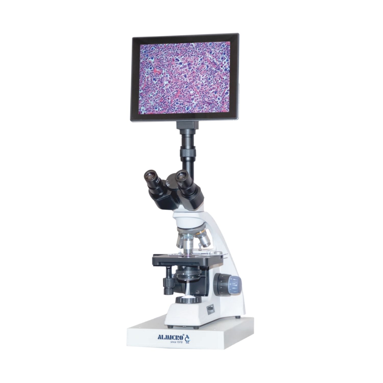
| Model | DVM-02 Plus |
| Head | Trinocular, 30° inclined, 360° rotatable, interpupillary 54–75 mm, diopter ±5 |
| Eyepiece | Wide Field WF10X/18mm |
| Objectives | Achromatic 4X, 10X, 40Xs, 100Xs (oil), quadruple nosepiece |
| Condenser | Abbe N.A. 1.25, iris diaphragm with filter holder |
| Focusing | Coaxial coarse & fine, fine 0.002 mm, coarse stroke 36 mm, adjustable rack stop, friction control |
| Stage | Double-layer mechanical, 110X125 mm, travel 30X75 mm |
| Illumination | 3W LED, brightness adjustable, powered 110–240V |
| Camera Sensor | 1/2.5’’ 5 MP color CMOS, 2592 x 1944 pixels, dynamic range 66.5 dB |
| Camera Frame Rate | 1280×720 @ 15 fps, 640×480 @ 30 fps |
| Tablet | 9.7-inch IPS LCD 1024×768, 1GB RAM, 2GB ROM, Android 4.2.2 |
| Software | Built-in measurement & particle analysis, HDMI output, SD card, Bluetooth |
| Power | DC 5V |
Key Features
• Trinocular head inclined at 30° and 360° rotatable
• Wide-field eyepiece WF10X/18mm for comfortable viewing
• Quadruple achromatic objectives: 4X, 10X, 40Xs, 100Xs (oil)
• Coaxial coarse and fine focusing with 0.002 mm fine division
• Adjustable rack stop and friction control for sample protection
• Double-layer mechanical stage 110X125 mm with 30X75 mm travel
• Abbe condenser with iris diaphragm and filter holder
• 3W LED illumination with adjustable brightness
• 5 MP CMOS camera with 2592 x 1944 pixel resolution
• Android 9.7-inch tablet with built-in microscopy software
• Supports measurement, particle analysis, HDMI output, SD card storage, and Bluetooth
• Compact, portable, and easy-to-use integrated digital system
Working Principle
The DVM-02 Plus operates by directing light through the specimen on the mechanical stage. The objectives magnify the image, which is captured by the 5 MP CMOS camera. The image is displayed on the integrated Android tablet in real-time. Users can adjust focus, LED brightness, and stage position to get a sharp image. The tablet software allows measurement, particle analysis, and image storage, making observation and analysis seamless.
Use Cases
• Educational laboratories for teaching and demonstrations
• Research labs for biological or material analysis
• Industrial inspection for particle and surface analysis
• Quality control in manufacturing and electronics
• Fieldwork requiring portable digital microscopy
• Student training for digital imaging and analysis

Reviews
There are no reviews yet.