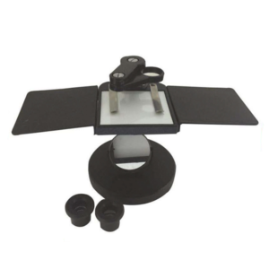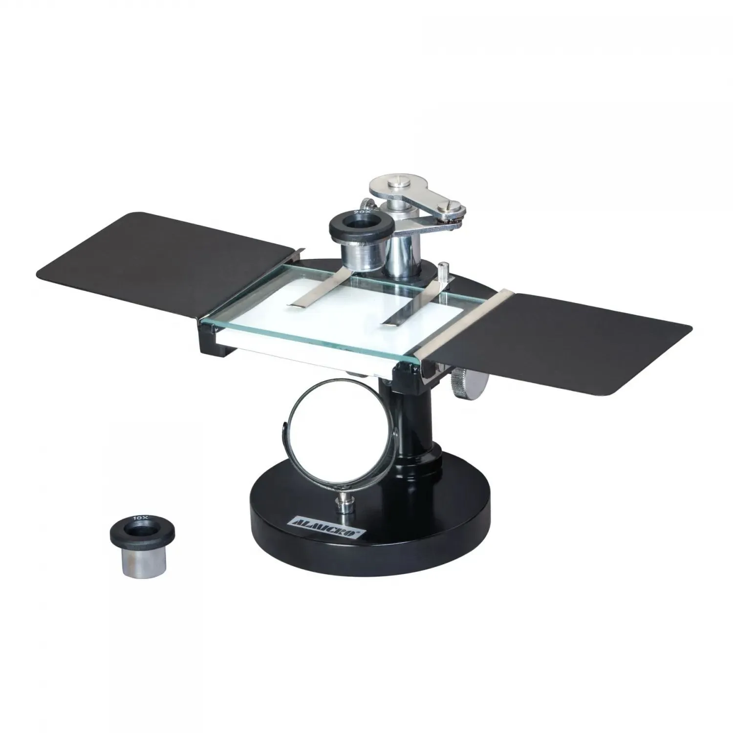
Description
Technical Specifications – Dissecting Microscope MD-1
Optical System
-
High-eye-point 10X eyepiece
-
High-eye-point 20X eyepiece
-
Crisp, wide field viewing suitable for long observation periods
Viewing Assembly
-
Jointed viewing arm
-
Enables full-area scanning of the stage
-
Easily adjustable for comfort and angle
-
Stage & Working Platform
-
Stage size: 85 mm × 75 mm
-
Glass stage with spring clips for specimen holding
-
White/black reversible metal plate for background contrast enhancement
-
Stable structure for detailed work
Illumination
-
Substage plano-concave mirror
-
Reflects natural daylight and artificial light effectively
-
Delivers uniform illumination across the specimen
Mechanical System
-
Rack & pinion mechanism enclosed inside the stage pillar
-
Ensures smooth vertical movement and long-term durability
Focusing System
-
Side-mounted focusing knobs
-
Provides smooth, precise, and controlled focusing action
User Comfort
-
Detachable hand rests on both sides for steady hand positioning
-
Reduces fatigue during detailed dissection or prolonged sessions
Use Case
-
Ideal for demonstration
-
Organism study
-
Biology training
-
Fine dissection work
-
Suitable for academic labs, research units, and field observation
Key Features
-
High-Clarity Optics: Equipped with high-eye-point 10X and 20X eyepieces for bright, sharp, and wide-field viewing.
-
Ergonomic Design: Jointed viewing arm ensures flexible positioning and complete stage coverage.
-
Dual-lighting compatibility: Substage plano-concave mirror supports both daylight and artificial light reflection.
-
Stable Working Platform: Large 85 × 75 mm stage with spring clips for secure specimen placement.
-
Enhanced Contrast Options: Comes with a white/black reversible plate and a glass stage for versatile background contrast.
-
Smooth & Precise Focusing: Side-mounted knobs ensure effortless, controlled focusing during delicate dissection.
-
Supportive Work Comfort: Detachable hand rests on both sides help reduce fatigue during prolonged use.
-
Ideal for Training & Demonstration: Perfect for biology students, teachers, and lab instructors for clear specimen study and fine dissection tasks.
Working Principle
The Dissecting Microscope MD-1 works on the principle of reflected light magnification. Unlike compound microscopes that use transmitted light, the MD-1 illuminates the specimen from above or by reflecting external light through a plano-concave mirror. The reflected light reaches the specimen surface and forms a magnified image when viewed through low-power eyepieces.
The high-eye-point eyepieces provide a wide field, making it easier to study three-dimensional specimens like insects, plant parts, fibers, crystals, or biological tissues. The rack-and-pinion mechanism adjusts the distance between the objective lens and the specimen, allowing the user to achieve sharp focus. The jointed arm ensures the observer can scan any part of the specimen without repositioning it, enabling smooth and continuous study.