Description
Technical Specification Table
| Specification | Details |
|---|---|
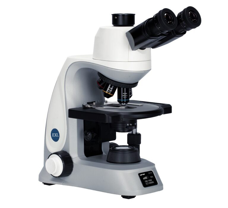
| Model | Almicro EXL Series Research Microscope |
| Frame | Modular upright, high-stability |
| Imaging | Brightfield and Fluorescence (upgradeable to Darkfield, DIC, Phase Contrast) |
| Illumination | 5W white LED transmitted light with Fly-Eye lens |
| Illumination Control | Auto-adjust based on objective, sleep mode included |
| Objectives | Plan Achromat 4X, 10X; Plan Fluor 20X, 40X, 100X |
| Nosepiece | Reversed sextuple with analyzer slot, smart recognition |
| Viewing Tube | Trinocular, 25 mm FOV, adjustable light path (100:0 / 50:50 / 0:100) |
| Eyepieces | High-eyepoint 10X, diopter adjustable |
| Stage | Coaxial mechanical stage, double slide holder |
| Condenser | Universal, high-contrast for Brightfield & Fluorescence |
| Fluorescence Module | Turret-type, 4 filter cubes, LED illumination, DAPI, FITC, TRITC |
Key Features
• Modular upright frame for Brightfield and Fluorescence imaging
• Fully upgradeable to Darkfield, DIC, Phase Contrast, and multi-head teaching
• Integrated system components for optimal performance and compatibility
• On-body status display for real-time system and illumination information
• High-performance 5W white LED transmitted illumination with Fly-Eye lens
• Smart illumination control adjusts light based on objective magnification
• Sleep mode for auto shut-off and energy conservation
• Reversed sextuple nosepiece for 6 objectives with analyzer slot
• Smart objective recognition and real-time scale bar calibration
• Trinocular viewing tube with wide 25 mm field of view and diopter adjustment
• Smooth mechanical stage with double slide holder
• Universal condenser optimized for Brightfield and Fluorescence
• Turret-type Fluorescence module with four filter cubes and LED illumination
Working Principle
Light from the high-performance LED passes through the specimen on the mechanical stage. Objectives collect and magnify the image, which is viewed through the trinocular eyepieces or captured via a camera. The system automatically adjusts illumination based on objective magnification. Fluorescence modules excite specific dyes in the sample, producing high-contrast images. Users control focus, illumination, and filters to acquire precise, clear images for analysis.
Use Cases
• Advanced biomedical research and cell biology studies
• Life sciences labs for Brightfield and Fluorescence imaging
• Materials science labs for structural and compositional analysis
• Educational institutions for multi-head teaching and training
• Research centers requiring upgradeable, modular imaging systems
• Digital imaging and microphotography for documentation and analysis
Reviews
There are no reviews yet.