Description
Technical Specification Table
| Specification | Details |
|---|---|
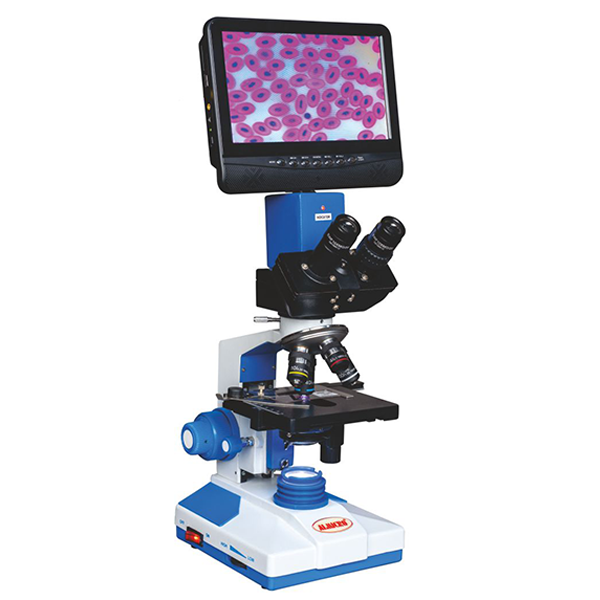
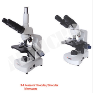
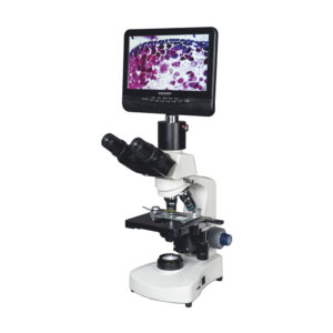
| Viewing Head | Compensation free Trinocular Head, inclined at 45° |
| Interpupillary Distance | 55–75 mm |
| Eyepiece (Ocular) | 10x (paired), field of view 20 mm |
| Objective | Achromatic: 4x, 10x, 40x, 100x |
| Nosepiece | Quadruple nosepiece with positive click stops; moves on ball bearings |
| Focusing System | Co-axial coarse and fine focusing, sensitivity 0.004 mm, range 24 mm |
| Condenser | Abbe N.A. 1.2 |
| Stage | Double layer mechanical stage, 132×142 mm; movement range 75×40 mm |
| Lamp House | LED Lamp, 3W |
| Display | 7″ TFT LED Screen, all functions controlled via remote |
| Valid Pixel | 3 MP |
| Power | DC 12V |
| Packaging | Inner thermocol/styrofoam packing and outer cardboard box |
Key Features
Trinocular head with 45° inclination for comfortable observation.
Interpupillary distance adjustable between 55–75 mm.
High-precision achromatic objectives: 4x, 10x, 40x, 100x.
Quadruple nosepiece with positive click stops.
Coaxial coarse and fine focusing system with 0.004 mm sensitivity.
Abbe condenser with N.A. 1.2 ensures clear illumination.
Double-layer mechanical stage with precise movement (75×40 mm).
LED lamp (3W) for low-heat illumination.
7″ LED display with 3MP resolution; easy remote control.
Robust build ideal for labs, classrooms, and research facilities.
Working Principle
The DVM-01 works by directing light through the specimen using an LED illumination system. The condenser and objectives magnify the sample, and users can view the image on the trinocular head or the 7″ LED display. Coarse and fine focus knobs adjust the stage for sharp clarity. Digital output allows image capture and recording via SD card.
Use Cases
Biology labs in universities and schools for teaching and demonstration.
Research laboratories for cellular, tissue, and microorganism studies.
Clinical and diagnostic labs for sample analysis.
Industrial labs for material and quality testing.
Training centers for collaborative microscopy and presentations.
Reviews
There are no reviews yet.