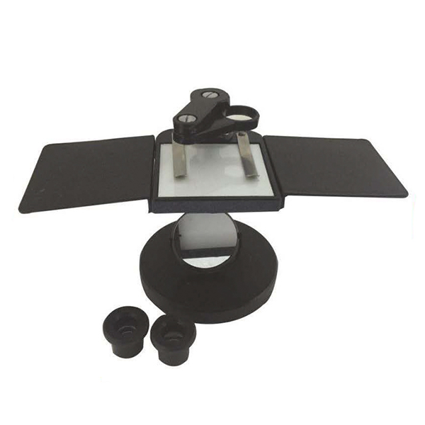
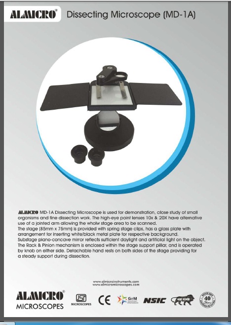
Description
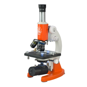
Technical Specifications – Dissecting Microscope MD-1A
| Specification | Details |
|---|---|
| Eyepieces | High-eyepoint 10X and 20X lenses |
| Viewing Arm | Jointed arm for full-stage scanning |
| Stage Size | 85 mm × 75 mm |
| Stage Design | Glass plate with spring clips; white/black background metal plate |
| Illumination | Substage plano-concave mirror for daylight and artificial light |
| Mechanism | Rack and pinion enclosed in stage support pillar |
| Focusing | Side-mounted focusing knobs |
| Hand Rests | Detachable hand rests on both sides |
| Use Case | Demonstration, organism study, fine dissection work |
Key Features
-
High-eyepoint 10X and 20X eyepieces for wide, bright, and comfortable viewing
-
Jointed viewing arm for scanning the entire stage area
-
Stable 85 × 75 mm stage with spring clips for secure specimen placement
-
Glass stage plate with white/black reversible metal plate for contrast
-
Substage plano-concave mirror for effective daylight and artificial light reflection
-
Rack and pinion mechanism enclosed in the support pillar for durability
-
Smooth side-mounted knobs for precise focusing
-
Detachable hand rests for steady dissection and reduced fatigue
-
Ideal design for live specimen study and classroom demonstrations
-
Robust construction suitable for long-term laboratory use
Working Principle
The Dissecting Microscope MD-1A works on a reflected light system. Light from the environment or an external source is directed onto the specimen using a plano-concave mirror. The reflected light illuminates the specimen’s surface and forms a magnified image through low-power eyepieces.
The high-eyepoint eyepieces allow comfortable viewing of three-dimensional objects such as insects, plant tissues, small organisms, and surface structures. The rack and pinion system adjusts the vertical distance between the specimen and the optical components, helping the user achieve a sharp and clear focus.
The jointed viewing arm lets the observer scan the entire stage without moving the specimen, making observation continuous and smooth.
Use Cases
-
Biology classroom demonstrations
-
Study of insects, larvae, plant parts, and small organisms
-
Fine dissection work in educational labs
-
Training and practical teaching sessions
-
Field sample observation and basic research tasks
Reviews
There are no reviews yet.