Description
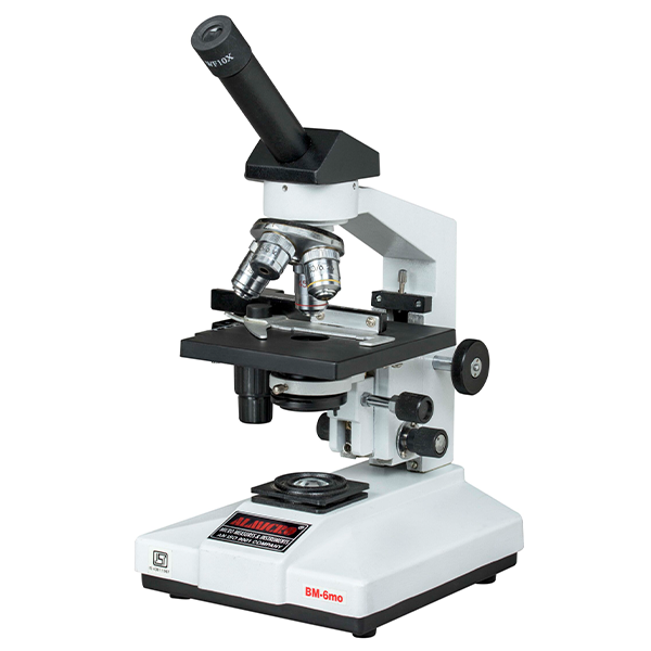
BM–6MO Microscope – Technical Specifications & Features
Technical Specifications
| Specification | Details |
|---|---|
| Head Type | 45° inclined monocular for ergonomic viewing |
| Objectives | Achromatic objectives for sharp and color-corrected imaging |
| Condenser | Abbe condenser NA 1.25 with iris diaphragm for precise light control |
| Focusing | Coaxial coarse and fine focusing system for smooth adjustments |
| Illumination | LED illumination for bright, uniform light |
| Stage | Sturdy mechanical stage for stable slide placement |
| Usage | Suitable for pathology, biology, and clinical laboratories |
Key Features
45° inclined monocular head ensures comfortable and strain-free observation.
Achromatic objectives deliver sharp, color-accurate magnification.
Abbe condenser (NA 1.25) with iris diaphragm allows optimal control over light and contrast.
Coaxial coarse and fine focusing system enables precise focusing with smooth operation.
LED illumination provides bright, consistent, and energy-efficient lighting.
Durable mechanical stage keeps slides secure during examination.
Ergonomically designed for long laboratory sessions.
Perfect for pathology, biology, and clinical laboratories.
Produces high-contrast, bright images for accurate analysis and study.
Working Principle
The BM–6MO microscope works by directing LED light through the Abbe condenser onto the specimen. The achromatic objectives magnify the sample, while the inclined monocular head ensures comfortable viewing. Users can adjust focus precisely using the coaxial coarse and fine focusing knobs, while the mechanical stage allows smooth slide positioning. The iris diaphragm controls illumination and contrast, resulting in bright, clear, and high-contrast images, ideal for laboratory observation, research, and diagnosis.
Applications / Use Cases
Pathology Laboratories: Tissue and cell examination for accurate diagnosis.
Biology Labs: Teaching, research, and detailed biological studies.
Clinical Laboratories: Observation of diagnostic samples.
Educational Institutions: Microscopy training for students.
Research Facilities: Detailed microscopic studies for experiments and analysis.
Reviews
There are no reviews yet.