Description
Technical Specification Table
| Specification | Details |
|---|---|
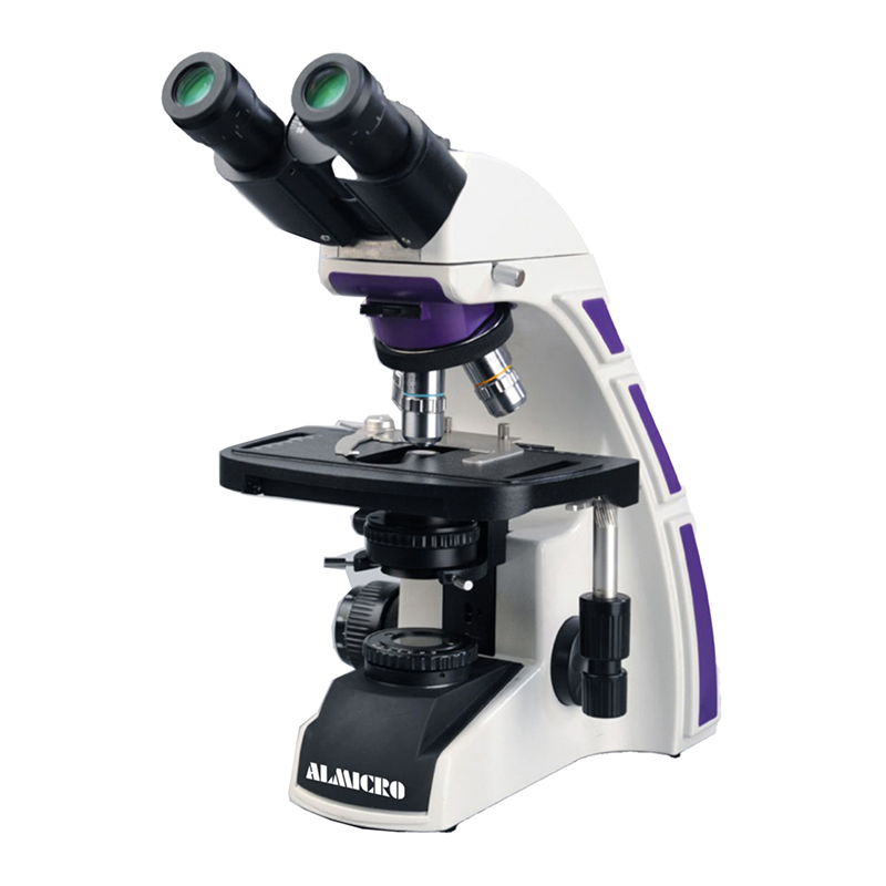
| Observation Tube | Binocular, articulated viewing head, 30° inclined |
| Interpupillary Distance | Adjustable, 50–75 mm |
| Eyepiece | PL10× / 23 mm high-eyepoint eyepieces |
| Diopter Adjustment | ±5 on left eyepiece |
| Optical System | Infinity-corrected finite optical system |
| Nosepiece | Backward-facing quintuple revolving nosepiece |
| Objective Lenses | Plan Achromatic 4×, 10×, 40× (Spring), 100× (Oil) |
| Mechanical Stage | Double-layer mechanical stage, 220 × 148 mm |
| Stage Travel Range | 76 × 50 mm with precision slide clamp |
| Condenser | Height-adjustable Abbe condenser, NA 1.25 |
| Iris Diaphragm | Integrated with filter holder |
| Focusing Mechanism | Coaxial coarse and fine focusing |
| Fine Focus Precision | 0.002 mm |
| Illumination | 3W LED with Koehler illumination |
| Light Control | Intensity adjustable |
| Power Supply | 230V AC, 50/60 Hz |
| Body Construction | Integrated metal body with ergonomic design |
| Packaging | Inner thermocol packing with outer corrugated box |
Key Features
-
Infinity-corrected optical system for superior resolution
-
Ergonomic binocular head reduces operator fatigue
-
High-quality Plan Achromatic objectives for distortion-free images
-
Precision coaxial coarse and fine focusing mechanism
-
Large double-layer mechanical stage for stable specimen handling
-
Adjustable Abbe condenser for uniform illumination
-
3W LED illumination with long operational life
-
Backward-facing quintuple nosepiece for smooth objective switching
-
Rugged metal body ensures vibration-free performance
-
Suitable for research, pathology, education, and clinical labs
Working Principle
The microscope operates on the principle of bright-field optical microscopy. Light generated from the 3W LED source passes through the condenser, which focuses illumination evenly onto the specimen. The specimen modulates the transmitted light, which is then magnified by the objective lens. The infinity-corrected optical system transmits parallel light rays to the eyepieces, producing a sharp, high-contrast image. Coaxial focusing allows precise control of specimen depth and clarity.
Case Study: Research Laboratory
A university life-science research laboratory integrated the ALMICRO VXL-BINO Microscope for cellular morphology studies and routine histology analysis. The lab required high optical clarity, stable mechanical performance, and long-hour usability. After installation, researchers reported improved image sharpness at higher magnifications and reduced eye strain due to the ergonomic binocular head. The LED illumination minimized maintenance costs, while the precision stage improved sample positioning accuracy, enhancing overall research productivity.
Reviews
There are no reviews yet.